American Scientists Discover Lower White Matter Cerebral Blood Flow In Those Who Have Recovered From Mild COVID-19!
COVID-19-News - Lower White Matter -Cerebral Blood Flow - Mild-COVID-19 Jun 04, 2023 2 years, 6 months, 3 weeks, 6 days, 5 hours, 7 minutes ago
Groundbreaking Study Reveals Link Between Mild COVID-19 and Changes in Brain Blood Flow
COVID-19 News: In a study conducted at the University of South Carolina, scientists have made a significant breakthrough in understanding the impact of COVID-19 on the brain. The research reveals that even mild cases of COVID-19 can lead to lower white matter cerebral blood flow, shedding light on the potential long-term consequences of the disease. These study findings challenge the notion and fallacies including previous
COVID-19 News reports that claim mild cases of COVID-19 result in complete recovery and emphasize the importance of further investigation into the effects of the virus on brain health.
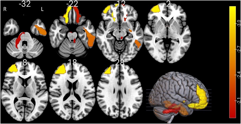
 Areas where the COVID-19 group had significantly lower cerebral blood flow than the control group. White numbers indicate
axial slice location. The color bar represents the z-scores for this contrast. Only statistically significant regions are shown.
Data are derived from univariate analysis. R, right; L, left.
The Study
The study team compared a group of individuals who had recovered from mild COVID-19 with a control group of healthy individuals. Using advanced brain imaging techniques, they measured cerebral blood flow (CBF) in different regions of the brain, including gray matter (GM) and white matter (WM). The results were striking: both whole-brain CBF and WM CBF were significantly lower in the COVID-19 group compared to the control group.
Implications and Potential Markers
The study's findings suggest that altered blood flow patterns in the brain could serve as an imaging marker for mild COVID-19 infection. Importantly, the research showed that predictive models based on these CBF patterns accurately identified COVID-19 patients, demonstrating a high degree of accuracy in determining group membership. This breakthrough opens up new possibilities for using brain imaging as a diagnostic tool for COVID-19, potentially allowing for early identification and intervention in patients.
Understanding the Effects of Mild COVID-19
While severe cases of COVID-19 have received significant attention due to their devastating impact, mild cases make up the majority of infections and can still have long-term consequences. Survivors of mild-to-moderate COVID-19 may experience cognitive, behavioral, or health-related outcomes that require further investigation. Previous studies have primarily focused on severe cases, making this research significant in highlighting the potential brain changes associated with mild COVID-19.
The Link Between Blood Flow and Neurological Manifestations
The observed alterations in blood flow patterns corresponded to specific brain areas and white matter tracts are known to be involved in various neurological functions. Changes in the basal ganglia and cerebral peduncles were linked to balance and dizziness issues reported
by COVID-19 patients, while alterations in the inferior frontal and olfactory tubercle were associated with changes in the sense of smell. Additionally, changes in the frontal and temporal lobes and anterior cingulate, responsible for executive function and language processing, could explain the reported "brain fog" and speech processing difficulties in COVID-19 patients.
Potential for Early Detection and Long-Term Prognosis
Remarkably, the study's predictive models showed promising results in accurately discriminating between individuals with and without COVID-19. By leveraging brain-based approaches, researchers achieved an unprecedented level of accuracy in identifying COVID-19 infections. This breakthrough discovery opens doors for the development of larger-scale studies and longitudinal research to better understand the long-term neurological consequences of COVID-19 and predict recovery outcomes based on CBF patterns.
Next Steps and the Road Ahead
While the study sheds light on the connection between mild COVID-19 and changes in cerebral blood flow, the underlying physiological mechanisms require further investigation. Understanding the relationship between vascular function and COVID-19 infection is essential for unraveling the complexities of the disease.
Future research should explore the potential associations between inflammatory markers, disease severity, and cerebral blood flow changes. Furthermore, ongoing studies should investigate how alterations in CBF relate to changes in white matter tracts and connectivity in COVID-19 patients.
Conclusion
The University of South Carolina study marks a significant step forward in unraveling the mysteries surrounding COVID-19 and its impact on the brain. The discovery of lower white matter cerebral blood flow in patients recovered from mild COVID-19 emphasizes the importance of understanding the long-term consequences of the disease, even in cases that were initially considered mild.
The study findings have far-reaching implications for healthcare professionals, researchers, and policymakers. By identifying a potential imaging marker for COVID-19 infection, healthcare providers may be able to diagnose the disease more accurately and intervene earlier, leading to improved patient outcomes. Moreover, this breakthrough paves the way for further investigations into the mechanisms underlying cerebral blood flow changes and their relationship to cognitive and behavioral outcomes.
The future holds great promise for additional research and studies that build upon these findings. Longitudinal studies can assess the long-term effects of altered cerebral blood flow and determine whether it is associated with cognitive decline, chronic fatigue, and other persistent symptoms reported by COVID-19 survivors.
The study findings were published in the peer reviewed Journal of Neuroimaging.
https://onlinelibrary.wiley.com/doi/10.1111/jon.13129
For the latest
COVID-19 News, keep on logging to Thailand Medical News.
