Nikhil Prasad Fact checked by:Thailand Medical News Team Dec 03, 2024 4 months, 3 weeks, 2 days, 1 hour, 30 minutes ago
Medical News: Axon Shape Redefined by New Research
For more than a century, biology textbooks have described axons - key structures in brain cells - as cylindrical tubes with consistent diameters. These depictions have shaped the way we understand how brain cells communicate and function. However, a groundbreaking study by researchers from Johns Hopkins University School of Medicine and University of California, San Diego School of Medicine challenges this long-standing view. Their findings suggest that axons are far more dynamic than previously believed, resembling a "pearls-on-a-string" structure rather than uniform tubes.
 Axon Shape of Brain Cells May Not Be What We Thought
This new understanding has far-reaching implications for neuroscience, influencing how we perceive brain cell communication, plasticity, and the development of neurodegenerative diseases. This
Medical News report delves into the details of this transformative study, its findings, and its implications for human health.
A Century-Old Assumption
Axons are the armlike structures of neurons that connect brain cells and facilitate the transmission of electrical signals. These structures are essential for brain functions like learning, memory, and sensory processing. Traditional illustrations and models have portrayed axons as cylindrical tubes with a consistent diameter, occasionally interrupted by synaptic structures where neurotransmitters are exchanged.
The study, led by Dr. Shigeki Watanabe at Johns Hopkins and Dr. Padmini Rangamani at University of California, San Diego, challenges this long-held assumption. Instead of smooth tubes, the researchers observed a structure resembling "pearls-on-a-string" under near-physiological conditions.
Using advanced high-pressure freezing electron microscopy, the team preserved the natural shape of axons by freezing them quickly. This method prevented distortions caused by conventional preparation techniques like chemical dehydration. The team analyzed axons in three types of mouse neurons: neurons grown in laboratory conditions, neurons from adult mice, and neurons from mouse embryos. In all cases, they found bead-like structures - dubbed “non-synaptic varicosities” - along the length of the axons.
Why Axonal Shape Matters
The discovery of the pearls-on-a-string morphology has significant implications for our understanding of brain function. Axons are not merely passive conduits for transmitting electrical signals; their shape actively influences how these signals are conducted.
Dr. Watanabe explains, "The physical properties of axons affect how ions move through them. Wider spaces in axons allow ions to pass more efficiently, reducing delays and traffic jams. This structural variation could influence how the brain processes information and responds to changes in its environmen
t."
Interestingly, this type of pearling structure has been observed before in neurons undergoing degeneration, such as in neurodegenerative conditions like Parkinson’s disease. However, this study reveals that these structures are also present in healthy axons, suggesting they play a functional role rather than solely indicating damage.
Investigating the Cause of Pearling
The researchers sought to uncover what drives the formation of this unique structure. They found that three key factors contribute to the pearls-on-a-string morphology:
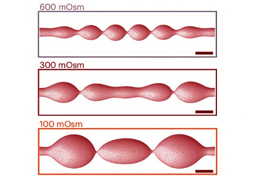
-Membrane Tension: The tension in the axonal membrane significantly affects its shape. When the researchers reduced membrane tension by altering sugar concentrations in the surrounding solution, the pearls became smaller.
-Cholesterol Levels: Cholesterol is crucial for maintaining membrane stiffness. By removing cholesterol, the team observed that the axonal membrane became more fluid-like, and the pearl structures diminished. However, this also impaired the axon’s ability to efficiently transmit electrical signals.
-Neuronal Activity: When neurons were subjected to high-frequency electrical stimulation, the pearls along the axons temporarily swelled, becoming 8% longer and 17% wider. This swelling persisted for at least 30 minutes, indicating that axonal shape adapts dynamically to activity levels.
Advanced Techniques and Mathematical Modeling
To study axonal structure at such a fine level, the team employed high-pressure freezing electron microscopy. Unlike traditional methods that dehydrate tissues, this technique freezes axons rapidly, preserving their natural shape. Dr. Watanabe likened this process to freezing a grape instead of drying it into a raisin, ensuring that the axon's structure remains intact.
Additionally, the researchers collaborated with mathematical modeling experts to simulate the physical forces acting on axons. Their models showed that the pearl-like structures could be explained by simple mechanical principles, such as the balance of membrane tension and pressure. These simulations matched their experimental observations, providing a comprehensive picture of how axonal shapes are formed and maintained.
Implications for Neurodegenerative Diseases
One of the most exciting aspects of this research is its potential implications for neurodegenerative diseases. Pearl-like structures in axons have been associated with dying neurons in conditions like Alzheimer’s and Parkinson’s disease. However, the discovery that healthy axons also exhibit this morphology suggests that changes in the pearls-on-a-string structure might serve as early indicators of disease.
The research team plans to extend their work to human brain tissue, including samples from individuals with neurodegenerative conditions. This could reveal whether abnormal pearling patterns are linked to disease progression or whether interventions targeting axonal structure could slow or prevent neurodegeneration.
Biophysical Forces at Play
The study highlights the importance of biophysical forces in shaping axons. The researchers found that:
-Osmotic Pressure: Increasing the concentration of sugars around axons caused the pearls to shrink, while decreasing sugar concentrations made the pearls larger.
-Cholesterol Removal: Reducing cholesterol levels in the membrane made the axons less stiff and decreased pearling. However, this also reduced the speed of electrical signals, suggesting a trade-off between structural flexibility and functional efficiency.
-Electrical Stimulation: High-frequency stimulation not only increased the size of the pearls but also improved the speed of electrical signal transmission, showing that axons can adapt their shape to optimize performance.
These findings suggest that the pearls-on-a-string morphology is not static but highly dynamic, responding to both internal and external cues.
Conclusions
This study represents a paradigm shift in our understanding of axonal structure and function. Key conclusions include:
-Dynamic Morphology: Axons are not uniform tubes but dynamic structures with bead-like swellings.
-Role of Membrane Mechanics: The shape of axons is governed by the balance of membrane tension, cholesterol levels, and external stimuli.
-Functional Implications: The pearls-on-a-string structure influences how electrical signals travel through axons, potentially affecting learning, memory, and other cognitive processes.
-Disease Relevance: Changes in axonal morphology could serve as early markers of neurodegenerative diseases or targets for therapeutic intervention.
The study highlights the complexity of the brain at the microscopic level, showing that even seemingly minor structural details can have profound effects on function. As Dr. Watanabe notes, "Understanding the shape of axons opens new doors for exploring brain plasticity and developing treatments for neurological diseases."
The study findings were published in the peer-reviewed journal: Nature Neuroscience.
https://www.nature.com/articles/s41593-024-01813-1
For the latest Medical Discoveries, keep on logging to Thailand
Medical News.
Read Also:
https://www.thailandmedical.news/news/breaking-medical-news-international-medical-community-now-recognizes-the-aorta-as-an-independent-organ
https://www.thailandmedical.news/news/breaking-medical-news-international-study-shows-that-young-individuals-who-got-ct-scans-have-an-increased-risk-of-developing-cancer
