BREAKING! Brazil Study Provides Evidence That SARS-CoV-2 Infects The Astrocytes Of The Human Brain Using NRP1 Receptors And Causes Damage!
Source: Medical News - NeuroCOVID Aug 12, 2022 2 years, 8 months, 1 week, 6 days, 11 hours, 1 minute ago
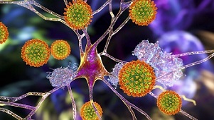
NeuroCOVID: A new study by researchers from Institute of Biology at the University of Campinas-Brazil has provided alarming evidence that the SARS-CoV-2 coronavirus infects astrocytes and to a lesser extent, neurons of the human brain by using the NRP1 (Neuropilin-1) receptors and in the process causes damage to the brain!

It has already widely known that neurological symptoms are among the most prevalent of the extrapulmonary complications of COVID-19, affecting more than 30% of patients.
The
NeuroCOVID study team provides evidence that severe acute respiratory syndrome coronavirus 2 (SARS-CoV-2) is found in the human brain, where it infects astrocytes and to a lesser extent, neurons.
The study findings also show that astrocytes are susceptible to SARS-CoV-2 infection through a noncanonical mechanism that involves spike–NRP1 interaction and respond to the infection by remodeling energy metabolism, which in turn, alters the levels of metabolites used to fuel neurons and support neurotransmitter synthesis. The altered secretory phenotype of infected astrocytes then impairs neuronal viability. These features could explain the damage and structural changes observed in the brains of COVID-19 patients.
Despite increasing evidence confirming neuropsychiatric manifestations associated mainly with severe COVID-19 infection, long-term neuropsychiatric dysfunction (recently characterized as part of “long COVID-19” syndrome) has been frequently observed even after mild infection.
The study findings show the spectrum of cerebral impact of severe acute respiratory syndrome coronavirus 2 (SARS-CoV-2) infection, ranging from long-term alterations in mildly infected individuals (orbitofrontal cortical atrophy, neurocognitive impairment, excessive fatigue and anxiety symptoms) to severe acute damage confirmed in brain tissue samples extracted from the orbitofrontal region (via endonasal transethmoidal access) from individuals who died of COVID-19.
In an independent cohort of 26 individuals who died of COVID-19, the study team used histopathological signs of brain damage as a guide for possible SARS-CoV-2 brain infection and found that among the 5 individuals who exhibited those signs, all of them had genetic material of the virus in the brain.
Importantly, brain tissue samples from these five patients also exhibited foci of SARS-CoV-2 infection and replication, particularly in astrocytes. Supporting the hypothesis of astrocyte infection, neural stem cell–derived human astrocytes in vitro are susceptible to SARS-CoV-2 infection through a noncanonical mechanism that involves spike–NRP1 interaction.
Interestingly, SARS-CoV-2-infected astrocytes manifested changes in energy metabolism and in key proteins and metabolites used to fuel neurons, as well as in the biogenesis of neurotransmitters. Moreover, human astrocyte infection elicits a secretory phenotype that reduces neuronal viability.
The study findings support the model in which SARS-CoV-2 reaches the brain, infects astrocytes, and consequently, leads to neuronal death or dysfunction. These deregulated processes could contribute to the structural and functional alterations seen in the brains of COVID-19 patients.
The study findings were published in the peer re
viewed journal: PNAS
https://www.pnas.org/doi/full/10.1073/pnas.2200960119
The study findings are even more worrisome as some of the newly emerging variants and subvariants ie the BA.5 variant and its various subvariants along with some of the newer BA.2 subvariants could be even more neuropathogenic as symptoms similar to encephalitis seems to become more pronounced in many infected with these newer emerging variants.
https://www.thailandmedical.news/news/breaking-sars-cov-2-ba-5-variant-could-be-more-neuropathogenic-urgent-studies-warranted-to-assess-long-covid-threats-to-the-brain-and-cns
Many infected with these newer variants could end up developing a variety of neurological issues in the long term.
The study findings demonstrate structural and functional alterations in the brain tissue of COVID-19 patients, which parallel in vivo findings of cortical atrophy, neuropsychiatric symptoms, and cognitive dysfunctions.
A recent longitudinal study with 401 individuals (median age of 62 y, infected between March 2020 and April 2021, scanned pre- and postinfection) reported atrophy in the orbitofrontal and parahippocampal regions and cognitive impairment (determined by Color Trail tests).
https://pubmed.ncbi.nlm.nih.gov/35255491/
The patients the Brazilian study team analyzed were infected between March and July 2020 (and therefore, were most likely infected with the original SARS-CoV-2 strain), and the study team also observed atrophy in the orbitofrontal area and cognitive dysfunction (longer time to perform Color Trail tests and poorer verbal memory task performance).
Interestingly, patients with only mild COVID-19 also exhibited cortical atrophy in the superior temporal gyrus, which was previously described in a group of patients with severe SARS-CoV-2 infection.
https://pubmed.ncbi.nlm.nih.gov/33630760/
The study team also observed that higher levels of anxiety symptoms correlated with atrophy of the orbitofrontal cortex, a region previously linked with anxiety disorders.
https://pubmed.ncbi.nlm.nih.gov/17698998/
The study findings suggest that anxiety and depression symptoms are at least partially associated with SARS-CoV-2 infection, a hypothesis supported by a recently discovered association between anxiety and reactive astrogliosis in patients after COVID-19.
https://nn.neurology.org/content/9/3/e1151.abstract
Study findings showing alterations in brain structure and the manifestation of neurological symptoms in COVID-19 patient raise a debate on whether these clinical features are a consequence of peripheral changes or rather, viral invasion of the CNS.
https://pubmed.ncbi.nlm.nih.gov/32298803/
https://pubmed.ncbi.nlm.nih.gov/32437679/
https://www.ncbi.nlm.nih.gov/pmc/articles/PMC8562542/
According to the Brazilian study team, both hypotheses are possible as they detected histopathological alterations associated with SARS-CoV-2 presence in brain tissue collected from 5 deceased patients, while 21 individuals who died of COVID-19 did not show any brain tissue alterations. However, as the sampling region was small, the possibility remains that other brain regions may exhibit COVID-19–related histopathological alterations. Indeed, the limited number of individuals who exhibited brain alterations associated with CNS SARS-CoV-2 detection and the imprecise and heterogenic nature of postmortem sample collection across studies may explain the discussion regarding the potential correlations between neuroinvasion and COVID-19 symptoms.
https://www.biorxiv.org/content/10.1101/2020.06.25.169946v2
https://pubmed.ncbi.nlm.nih.gov/32876341/
https://pubmed.ncbi.nlm.nih.gov/32753756/
https://pubmed.ncbi.nlm.nih.gov/33166988/
https://pubmed.ncbi.nlm.nih.gov/34244682/
https://pubmed.ncbi.nlm.nih.gov/34153974/
Though some studies failed to detect the virus in the CNS, others have found viral particles in the brain localized to the microvasculature and neurons, the choroid plexus, or meninges.
In vitro models, such as stem cell–derived neural cells and cerebral organoids, have also demonstrated that SARS-CoV-2 potentially infects brain cells.
However, the magnitude of the CNS infection, its distribution within the brain tissue, and the molecular and cellular bases underlying the phenomenon had not been thoroughly explored.
The Brazilian study team shows that astrocytes are the main site of infection and possibly, replication of SARS-CoV-2 in the brains of COVID-19 patients as evidenced by the detection of the viral genome, the SARS-CoV-2 spike protein, and dsRNA in postmortem brain tissue, ex vivo brain slices, and in vitro infected astrocytes.
The study findings corroborate past research that showed that astrocytes from primary human cortical tissue and stem cell–derived cortical organoids are susceptible to SARS-CoV-2 infection.
https://www.biorxiv.org/content/10.1101/2020.06.25.169946v2
https://pubmed.ncbi.nlm.nih.gov/34244682/
https://www.biorxiv.org/content/10.1101/2021.01.17.427024v1
A recent study described that SARS-CoV-2 could access the CNS through the neural–mucosal interface in olfactory mucosa, thereby entering the primary respiratory and cardiovascular control centers in the medulla oblongata.
https://pubmed.ncbi.nlm.nih.gov/33257876/
Other proposed routes of SARS-CoV-2 neuroinvasion include brain endothelial cells. Besides the inflammatory response produced by SARS-CoV-2 infection, endothelial cell infection could also cause dysfunctions in BBB integrity and facilitate further access of the virus to the brain.
https://pubmed.ncbi.nlm.nih.gov/34489403/
https://pubmed.ncbi.nlm.nih.gov/33113348/
https://pubmed.ncbi.nlm.nih.gov/33405097/
Even with the advances that have already been made, there is still much left to be learned about the routes that SARS-CoV-2 can take to invade the brain and how the virus navigates across different brain regions.
Although ACE2 is the best-characterized cellular receptor for SARS-CoV-2 to enter cells via interaction with the viral spike protein, other receptors have also been identified as mediators of infection.
From the current study findings and others, astrocytes do not express ACE2; rather, they exhibit elevated expression of NRP1, another SARS-CoV-2 spike target that is abundantly expressed in the CNS, particularly in astrocytes.
https://pubmed.ncbi.nlm.nih.gov/33031735/
https://pubmed.ncbi.nlm.nih.gov/33082294/
https://pubmed.ncbi.nlm.nih.gov/33082293/
https://pubmed.ncbi.nlm.nih.gov/33450186/
Importantly when NRP1 is blocked with neutralizing antibodies, SARS-CoV-2 infection in these cells is greatly reduced. These results indicate that SARS-CoV-2 infects in vitro astrocytes via the NRP1 receptor, although this has yet to be confirmed in vivo.
.jpg) SARS-CoV-2 infects the CNS, replicates in astrocytes, and causes brain damage. (A) Histopathological H&E images of postmortem brain tissue from individuals who died of COVID-19. Five of 26 individuals showed signs of brain damage as represented in the images by (A, I) areas of necrosis, cytopathic damage (i.e., enlarged, hyperchromatic, atypical-appearing nuclei), (A, II) vessels with margination of leukocytes and thrombi, and (A, III) an infiltration of immune cells. The alterations are indicated by red asterisks and respective zoomed-in images (Lower). Images were acquired with 400× magnification. (Scale bars: 50 µm.) (B) Viral load in brain tissues from the five COVID-19 patients who manifested histopathological alterations in the brain as compared with samples from SARS-CoV-2–negative controls (n = 5 per group). *P < 0.05 compared with the control group. (C) Representative confocal images of the brain tissue of one COVID-19 patient who manifested histopathological alterations. Immunofluorescence targeting GFAP (red), dsRNA (magenta), SARS-CoV-2-S (green), and nuclei (DAPI; blue). Images were acquired with 630× magnification. (Scale bars: 50 µm.) (D) Percentage of SARS-CoV-2-S–positive cells in the tissue of the five COVID-19 patients. (E) Percentage of GFAP+ vs. unidentified cells, Iba1+, and NeuN+ among all infected cells. Ten fields/cases were analyzed. (F) Cell type enrichment analysis using the dataset generated from postmortem brain tissue from patients who died of COVID-19. Dot size represents the number of proteins related to the respective cell type, and the color represents the P value adjusted by false discovery rate. All data are shown as mean ± SEM. P values were determined by two-tailed unpaired tests with Welch's correction (B) or ANOVA one way followed by Tukey’s post hoc test (E). H&E: hematoxylin and eosin; DAPI: 4′,6-diamidino-2-phenylindole.
SARS-CoV-2 infects the CNS, replicates in astrocytes, and causes brain damage. (A) Histopathological H&E images of postmortem brain tissue from individuals who died of COVID-19. Five of 26 individuals showed signs of brain damage as represented in the images by (A, I) areas of necrosis, cytopathic damage (i.e., enlarged, hyperchromatic, atypical-appearing nuclei), (A, II) vessels with margination of leukocytes and thrombi, and (A, III) an infiltration of immune cells. The alterations are indicated by red asterisks and respective zoomed-in images (Lower). Images were acquired with 400× magnification. (Scale bars: 50 µm.) (B) Viral load in brain tissues from the five COVID-19 patients who manifested histopathological alterations in the brain as compared with samples from SARS-CoV-2–negative controls (n = 5 per group). *P < 0.05 compared with the control group. (C) Representative confocal images of the brain tissue of one COVID-19 patient who manifested histopathological alterations. Immunofluorescence targeting GFAP (red), dsRNA (magenta), SARS-CoV-2-S (green), and nuclei (DAPI; blue). Images were acquired with 630× magnification. (Scale bars: 50 µm.) (D) Percentage of SARS-CoV-2-S–positive cells in the tissue of the five COVID-19 patients. (E) Percentage of GFAP+ vs. unidentified cells, Iba1+, and NeuN+ among all infected cells. Ten fields/cases were analyzed. (F) Cell type enrichment analysis using the dataset generated from postmortem brain tissue from patients who died of COVID-19. Dot size represents the number of proteins related to the respective cell type, and the color represents the P value adjusted by false discovery rate. All data are shown as mean ± SEM. P values were determined by two-tailed unpaired tests with Welch's correction (B) or ANOVA one way followed by Tukey’s post hoc test (E). H&E: hematoxylin and eosin; DAPI: 4′,6-diamidino-2-phenylindole.
In order to understand the consequences of SARS-CoV-2 infection in NSC-derived astrocytes, the study team searched for changes in the proteome in a non-hypothesis-driven fashion. SARS-CoV-2 infection resulted in marked proteomic changes in several biological processes, including those associated with energy metabolism, in line with previous reports on other cell types infected with SARS-CoV-2.
https://pubmed.ncbi.nlm.nih.gov/33845483/
https://pubmed.ncbi.nlm.nih.gov/32619390/
Significantly, differentially expressed proteins in COVID-19 postmortem brains were enriched for astrocytic proteins more than oligodendrocytes, neurons, or Schwann cells, strengthening the findings that these are the most affected cells by SARS-CoV-2 infection in the human brain.
Corresponding author, Dr Daniel Martins-de-Souza from the Department of Biochemistry and Tissue Biology at the Institute of Biology-University of Campinas told Thailand
Medical News, “The study’s proteomic data also evidenced changes in the components of carbon metabolism pathways, particularly glucose metabolism, in both in vitro infected astrocytes and postmortem brain tissue from COVID-19 patients. As astrocyte metabolism is key to support neuronal function, changes in astrocyte metabolism could indirectly impact neurons.”
The study findings showed that that one of the most critical alterations caused by SARS-CoV-2 infection in astrocytes was a decrease in pyruvate and lactate levels.
Lactate exportation is one of the ways that astrocytes support neurons metabolically, shuttling this carbon source through the astrocyte–neuron lactate shuttle (ANLS) mechanism. In the ANLS, neuronal activity also enhances glutamate uptake by astrocytes. Lactate itself is essential for the support of neuronal activity and cerebral functions, acting as a neuroprotective agent as well as a key signal to regulate blood flow. These metabolic changes, particularly the reductions in lactate and pyruvate associated with decreases in MCT1 and MCT2 expression in SARS-CoV-2–infected astrocytes, support the hypothesis of ANLS disruption.
Furthermore, intermediates of glutamine metabolism, such as glutamate and GABA, were also decreased in SARS-CoV-2–infected astrocytes. This said, there were no significant changes in any core intermediates of the TCA cycle.
Alongside with an increased oxygen consumption rate of SARS-CoV-2–infected astrocytes, the study data suggest that glycolysis and glutaminolysis are being used to fuel carbons into the TCA cycle to sustain the increased oxidative metabolism of infected astrocytes.
A recent study showed that glutamine levels were also found reduced in mixed glial cells infected with SARS-CoV-2 and that the inhibition of glutaminolysis decreased viral replication and proinflammatory response, further reinforcing that glutamine could be used to fuel the TCA cycle in infected astrocytes.
https://www.biorxiv.org/content/10.1101/2021.10.23.465567v1
Although astrocyte-derived lactate is required for neuronal metabolism as previously mentioned, glutamine is used in the synthesis of neurotransmitters, such as glutamate and GABA.
Importantly astrocytes also play a vital role in glutamate-level homeostasis and neurotransmitter recycling, crucial processes for the maintenance of synaptic transmission and neuronal excitability. At glutamatergic synapses, glutamate uptake by astroglia prevents excitotoxicity, whereupon glutamine synthetase converts glutamate to glutamine, which can then be transferred back to neurons, thus closing the glutamate–glutamine cycle.
It was found that at the GABAergic synapses, GABA is taken up by astrocytes and first metabolized to glutamate before being converted to glutamine.
Considering the importance of the metabolic coupling between astrocytes and neurons, alterations in astrocytic glucose and glutamine metabolism are expected to compromise neuronal metabolism and plasticity and synaptic function.
Another past study using 18F-FDG PET analysis, reported hypometabolism in four different clusters of brain regions in patients suffering from long COVID-19, including the bilateral rectal/orbital gyrus and the olfactory gyrus.
https://pubmed.ncbi.nlm.nih.gov/33501506/
It has been found that as key regulators of CNS metabolism, alterations in astrocyte metabolism contribute to the 18F-FDG PET signal. Therefore, dysfunctions in astrocyte energy metabolism, like those observed here, could explain, at least partially, the brain hypometabolism in COVID-19 patients.
https://pubmed.ncbi.nlm.nih.gov/34966022/
https://pubmed.ncbi.nlm.nih.gov/34143277/
https://pubmed.ncbi.nlm.nih.gov/33813593/
https://pubmed.ncbi.nlm.nih.gov/33822001/
Besides the metabolic changes observed in SARS-CoV-2–infected astrocytes that may lead to neuronal dysfunction.
The study team found that SARS-CoV-2 infection elicits a neurotoxic secretory phenotype in astrocytes that results in increased neuronal death. A similar phenomenon has been observed when astrocytes are activated by inflammatory factors.
The alterations in cortical thickness the study team observed in COVID-19 patients could be explained by this neuronal death, at least partially, as well as by other mechanisms, including reactive astrogliosis and alterations in astrocyte specification and morphogenesis, as previously described in Alzheimer's disease, autism, and schizophrenia, as well as epilepsy, autism, and self-injury.
A past study reported astrogliosis in 86% of individuals who died following a diagnosis of SARS-CoV-2 infection.
https://pubmed.ncbi.nlm.nih.gov/33031735/
In agreement, snRNA-seq data show that the main markers of reactive astrocytes are enriched in samples from the medial frontal cortex of patients with COVID-19 compared with noninfected patients, supporting the hypothesis that reactive astrogliosis is a feature of COVID-19.
Astrocytes are also relevant in the regulation of synapses (and neural networks) and have been linked to the manifestation of depression, anxiety symptoms, and memory impairment, all of which have been observed in our post-COVID infection cohort.
The study findings are consistent with a model in which SARS-CoV-2 is able to reach the CNS of COVID-19 patients, infect astrocytes via NRP1 interaction, and secondarily impair neuronal function and viability. These changes may contribute to the alterations of brain structure observed here and elsewhere, thereby resulting in the neurocognitive and neuropsychiatric dysfunctions manifested by some patients with COVID-19.
The study findings come as a cautionary note that mechanisms of neuroinvasion in fatal COVID-19 could also be operative in mild COVID-19.
Importantly, interventions directed to treat COVID-19 should also envision ways to prevent SARS-CoV-2 invasion of the CNS and/or replication in astrocytes.
For the latest on
NeuroCOVID, keep on logging to Thailand
Medical News.

.jpg)