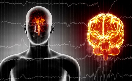
BREAKING COVID-19 News! Study Of EEG Signals Shows That Most Post-COVID Individuals Have Reduced Brain Activity!
Nikhil Prasad Fact checked by:Thailand Medical News Team Nov 26, 2023 1 year, 4 months, 4 days, 18 hours, 52 minutes ago
COVID-19 News: The COVID-19 pandemic, caused by the novel coronavirus SARS-CoV-2, has evolved into a global health crisis since its emergence in Wuhan, China, in late 2019. While primarily recognized for its severe respiratory manifestations, there is mounting evidence that COVID-19 can significantly affect the central nervous system, leading to neurological symptoms and complications.

A groundbreaking study covered in this
COVID-19 News report, conducted jointly by Tianjin First Central Hospital-China and Tianjin University-China has harnessed the power of electroencephalography (EEG) to delve into the intricate patterns of brain activity and connectivity in individuals recovering from COVID-19. This research aims not only to deepen our understanding of the neurological impacts of the virus but also to pave the way for advanced diagnostic and prognostic tools utilizing artificial intelligence.
The Neurological Landscape of COVID-19
As the COVID-19 pandemic continues to unfold, clinicians and researchers have increasingly observed neurological manifestations in infected individuals. These include headaches, dizziness, consciousness disorders, acute cerebrovascular disease, ataxia, epilepsy, and neuromuscular damage. Beyond clinical observations, experimental evidence points to changes in brain neurophysiological data, indicating that the virus might not be limited to respiratory effects.
Several studies have explored the changes in EEG characteristics in COVID-19 patients. Notably, alterations in EEG patterns and wave amplitudes have been reported, suggesting that the virus can impact brain activity directly. Moreover, distinct distribution patterns for EEG bands, higher Shannon’s spectral entropy, and altered hemispheric connectivity have been identified in COVID-19 patients, indicating potential damage to different brain regions. The causes of these EEG abnormalities are varied, including inflammatory damage, hypoxemia, and direct damage to brain neurons.
A study conducted on individuals recovering from COVID-19 revealed changing EEG characteristics over time, suggesting potential long-term effects on the central nervous system. However, existing research primarily focuses on changes in brain wave shapes, leaving the electrophysiological mechanism of nervous system injury in COVID-19 patients largely unexplored.
Objective and Methods of the EEG Study
The primary objective of this study was to investigate the abnormal levels of brain activity and changes in the brain's functional connectivity network in patients recovering from COVID-19, using EEG signals. The study employed a rigorous methodology, comparing and analyzing EEG signal sample entropy, energy spectrum, and brain network characteristic parameters in the delta (1–4 Hz), theta (4–8 Hz), alpha (8–13 Hz), and beta (13–30 Hz) bands.
The study involved 15 patients who had recovered from COVID-19 and 15 healthy controls. Electroencephalography signal sample entropy was used to calculate the complexity of EEG signals, offering insights into the activity levels of the two groups. Energy spectra were uti
lized to reflect the activity states of various brain regions. The directed transfer function (DTF) matrix, reflecting causal connection strength between cortical regions, was employed to construct a brain network model. Graph theory was then applied for quantitative analysis of the brain network, aiming to unravel the mechanism of the virus's impact on brain electrical activity.
Key Findings
The results of the study are both compelling and concerning. At rest, the energy values of the four frequency bands in the frontal and temporal lobes of COVID-19 patients were significantly reduced. Notably, specific leads, such as FP2, T3, and T4, showed a substantial decrease in energy values, indicating a widespread impact on diverse brain regions.
Furthermore, the study revealed a significant increase in sample entropy in the delta band of COVID-19 patients, suggesting heightened complexity in brain activity. In contrast, the beta band exhibited a significant decrease in sample entropy, indicating a potential reduction in attention, thinking activity, and cognitive flexibility in these patients.
While the average value of the directed transfer function did not show abnormalities, the study's network analysis demonstrated a significant rearrangement of core nodes in the brain functional network of COVID-19 patients. Specifically, the node degree in the temporal lobe increased, signifying enhanced mutual connections with other brain regions. In contrast, the input degree of the frontal and temporal lobes decreased, and the output degree of the frontal and occipital lobes increased, showcasing a profound reorganization of the core nodes in the brain network.
Discussion and Implications
The neurological impacts of COVID-19 extend beyond respiratory concerns, as evidenced by the findings of this EEG study. The observed reduction in energy levels in the frontal and temporal lobes aligns with previous research identifying brain cell reduction in areas controlling emotion and memory. This reduction in energy could contribute to cognitive, emotional regulation, and social cognitive impairments. Moreover, the increased complexity in the delta frequency band suggests more chaotic changes in brain activity, potentially indicating a heightened state of anxiety in COVID-19 patients.
The alterations in the beta frequency band, marked by decreased sample entropy, may further contribute to the understanding of the virus's impact on attention, thinking activity, and cognitive flexibility. The observed rearrangement of core nodes in the brain functional network emphasizes the need for a more comprehensive exploration of COVID-19's effects on brain connectivity and organization.
The study acknowledges certain limitations, including a small sample size and age differences between the control and patient groups. However, it underscores the feasibility of utilizing EEG signals to explore the neurophysiological impacts of COVID-19. Future research endeavors could focus on the dynamic changes in brain activity and network topology throughout the course of the disease, offering a more nuanced understanding of COVID-19's neurological effects.
Conclusion
In conclusion, the EEG study conducted by Tianjin First Central Hospital-China and Tianjin University-China unravels critical insights into the neurological impacts of COVID-19. The observed abnormalities in brain activity during rest, coupled with the reorganization of the brain functional network, provide a comprehensive understanding of the virus's impact on the nervous system. As the world grapples with the ongoing pandemic, these findings not only contribute to the scientific understanding of COVID-19 but also lay the foundation for future research, potentially enabling the application of artificial intelligence in diagnosing and predicting the prognosis of COVID-19 patients based on EEG signals. The study underscores the imperative for continued exploration into the multifaceted impacts of the virus on the central nervous system.
The study findings were published in the peer reviewed journal: Frontiers in Human Neuroscience.
https://www.frontiersin.org/articles/10.3389/fnhum.2023.1280362/full
For the latest
COVID-19 News, keep on logging to Thailand Medical News.
Read Also:
https://www.thailandmedical.news/news/breaking-covid-19-will-make-you-stupid-peer-reviewed-international-study-shows-that-sars-cov-2-infections-can-affect-one-s-intelligence
