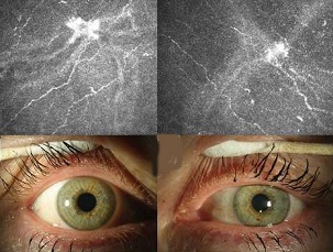
BREAKING COVID-19 News! Study Shows Most Long COVID Individuals Have Corneal Nerve Damage With 24.2 Percent Having Microneuromas!
Nikhil Prasad Fact checked by:Thailand Medical News Team Jan 27, 2024 1 year, 2 months, 5 days, 10 hours, 51 minutes ago
COVID-19 News: The enduring impact of COVID-19 extends beyond the acute phase, with a subset of individuals experiencing persistent symptoms, a phenomenon known as Long COVID-19 or post-COVID conditions. In a groundbreaking study covered in this
COVID-19 News report, conducted by researchers from the Complutense University of Madrid in Spain, corneal confocal microscopy has been employed to uncover a previously unnoticed facet of Long COVID-19 -corneal neuromas or microneuromas, and heightened immune activation, persisting over 20 months after SARS-CoV-2 infection.
 Most Long COVID Individuals Have Corneal Nerve
Damage With 24.2 Percent Having Microneuromas
Long COVID-19 - A Complex Continuation
Long COVID-19 has emerged as a multifaceted condition with symptoms ranging from fatigue, respiratory distress, to cognitive dysfunction. The Delphi consensus by the World Health Organization defines Long COVID-19 as symptoms persisting for at least two months, occurring within three months of a confirmed SARS-CoV-2 infection and without explanation by an alternative diagnosis. Recent studies have highlighted a negative impact on cognitive function, indicating linguistic–cognitive and visual attention impairment. The ongoing struggle with persistent symptoms significantly diminishes the quality of life and physical activity levels in individuals affected by Long COVID-19.
The Enigmatic Nervous System Connection
The neurological manifestations of COVID-19 have been diverse, encompassing headaches, fatigue, loss of taste and smell, brain fog, and neuropathic pain. Despite the prevalence of neurological symptoms, the precise mechanisms by which SARS-CoV-2 affects the nervous system remain unclear. However, studies suggest the involvement of both innate and adaptive immunity, with associations to small fiber neuropathy (SFN) and peripheral neuropathy.
Corneal Confocal Microscopy - A Window into Neuromas
In an effort to understand the impact of Long COVID-19 on the nervous system, the researchers conducted a cross-sectional comparative transversal study involving 53 participants - 33 with Long COVID-19 and 22 matched controls. The innovative use of corneal confocal microscopy allowed for a detailed examination of the sub-basal nerve plexus morphology.
The results revealed a significant alteration in corneal nerve parameters in Long COVID-19 patients. Reduced corneal nerve density, shorter corneal nerves, and lower branch density were observed compared to the control group. These findings, persisting over 20 months after acute SARS-CoV-2 infection, indicate a long-term impact on corneal innervation in individuals with Long COVID-19.
Dendritic Cells and Immune Activation
Dendritic cells (DCs), immune sentinels with the ability to bridge innate and adaptive immune responses, play a pivotal role in maintaining corneal nerve homeostasis. The study uncovered an intriguing aspect of immune activation in Long COVID-19 patients—heightened DC activa
tion. The areas occupied by DCs were notably larger in Long COVID-19 patients compared to the control group without systemic diseases. This immune response persisted even 20–24 months after the acute infection, suggesting a sustained inflammatory state.
Microneuromas - A Novel Discovery
One of the most striking revelations of the study was the detection of corneal microneuromas in 24.2% of Long COVID-19 patients. Also known as neuromas, these microscopic enlargements of terminal subbasal nerve endings are irregularly shaped and form at sites of nerve damage or injury. The presence of microneuromas indicates nerve regeneration following damage, a process observed in various pathological conditions. In the context of Long COVID-19, the microneuromas may signify the aftermath of nerve damage and subsequent attempt of recovery which may or may not be successful!
The Prolonged Impact of Long COVID-19
Comparisons with previous studies highlight the unique nature of this research. While some studies have reported a reduction in corneal nerve parameters in the aftermath of COVID-19 infection, this study goes a step further by demonstrating the persistent alterations in corneal innervation parameters even 20 months post-acute infection. The prolonged impact on corneal nerves, coupled with increased DC activation, underscores the chronic nature of the neuroinflammatory condition associated with Long COVID-19.
The Underlying Mechanism
The exact mechanism driving corneal nerve involvement in Long COVID-19 remains elusive. Inflammatory mediators and biochemical cascades are likely implicated in this condition. Neuropathological studies have shown the presence of SARS-CoV-2 in various neural tissues, accompanied by microglial activation and lymphoid inflammation. Additionally, COVID-19 infection has been associated with post-infectious immune-mediated peripheral neuropathy. The interplay of these factors may contribute to the persistent alterations observed in corneal innervation.
The Role of In Vivo Confocal Microscopy (IVCM)
The study's use of in vivo confocal microscopy (IVCM) as a noninvasive imaging technique provided a direct visualization of the corneal structure, including the sub-basal nerve plexus. IVCM has proven effective in identifying small nerve fiber damage in various peripheral neuropathies and immune-mediated SFN. While the cross-sectional nature of this study presents limitations, the potential for long-term follow-up and post-treatment assessments opens avenues for future investigations.
Conclusion and Future Directions
In conclusion, this comprehensive study illuminates the intricate connection between Long COVID-19 and corneal nerve damage. The persistent alterations in corneal nerve parameters, coupled with the presence of microneuromas and heightened DC activation, suggest a chronic neuroinflammatory condition in individuals with Long COVID-19, enduring over 20 months after acute SARS-CoV-2 infection.
As research continues to evolve, future studies should focus on long-term follow-ups to observe changes in corneal confocal findings and the potential impact of treatments. Addressing the inflammatory and immune responses observed in Long COVID-19 patients may lead to improved clinical management and better outcomes for those navigating the complex landscape of post-acute sequelae of SARS-CoV-2 infection.
The study was presented at the 2023 European Association for Vision and Eye Research Congress at Valencia in Spain. The study is still ongoing.
The study is available as abstract at the peer reviewed journal: Acta Ophthalmologica.
https://onlinelibrary.wiley.com/doi/10.1111/aos.16052
An earlier version of the initial study findings were published in the peer reviewed journal: Diagnostics.
https://www.mdpi.com/2075-4418/13/20/3188
An updated version is expected to be published soon at the time-line for follow ups reaches 36 months or 3 years but note that all figures stated in the studies are changing and in fact increasing!.... for instance the incidence of microneuromas are increasing!
For the latest
COVID-19 News, keep on logging to Thailand Medical News.
Read Also:
https://www.thailandmedical.news/news/breaking-covid-19-news-sars-cov-2-induced-retinal-microvascular-damage-is-not-reversible-many-will-eventually-become-blind
