BREAKING! COVID-19 Research: Scientist Discover Unusual Way SARS-CoV-2 Infects Other Cells Once Inside Human Host Without Need Of Receptors
Source: COVID-19 Research Aug 16, 2020 5 years, 4 months, 4 weeks, 1 day, 10 hours, 7 minutes ago
COVID-19 Research: Scientist from the University of California
, San Francisco found
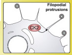
that when the SARS-CoV-2 virus infects human cells, the infected cell grows multi-pronged tentacles that are studded with viral particles. These filaments, called filopodia, reach out to still-healthy neighboring cells, which then bore into the cells’ bodies and infect the healthy cells with viral proteins.
.png)
This new discovery has phenomenal implications into how the virus is able to affect other tissues and organs in the human host without the need for receptors to bind to once inside the human host and also without the need for the full total virus to actual cause issues to these cells as the newly synthesized viral proteins are all that are needed to hijack the human host cells and body.

The research findings were published in the Journal: Cell
https://www.cell.com/cell/fulltext/S0092-8674(20)30811-4
Researchers previously believed the SARS-CoV-2 virus infected cells in a typical way, which is by finding receptors on the surface of cells in a person’s mouth, nose, respiratory tract, lungs or blood vessels, and replicating and invading larger cells.
This process of infiltration is not unique as other viruses, such as smallpox, HIV and some influenza viruses also used filopodia to improve their ability to infect cells.
The study showed that upon initial entry into human host cells using the various receptors including ACE2, the virus then hijack the host protein and kinase productions to start manufacturing its own proteins and enzymes and then using these it modifies certain host proteins involved in the cell cytoskeleton structures including ɑ-Catenin and Myosin IIa, to start creating cells that were laden with its own proteins and the filopodia structures to start infecting nearby cells.
.jpg) Colocalization of CK2 and Viral Proteins at Actin Protrusions
Colocalization of CK2 and Viral Proteins at Actin Protrusions
(A) Pathway of regulated PHs and SARS-CoV-2 interaction partners involved in cytoskeletal reorganization. Dashed lines indicate downregulation of activity, while solid lines indicate upregulation of activity. (B) Regulation of individual kinase activity or PHs depicted in (A).
(C) Caco-2 cells infected with SARS-CoV-2 at an MOI of 0.1 for 24 h prior to immunostaining for F-actin and M protein, as indicated. Shown is a confocal section revealing M protein localization along and to the tip of filopodia (left) and magnification of the dashed box (right).
(D) Dot plot quantification of the number and length of filopodia in untreated (mock) or infected Caco-2 cells for 24 h with SARS-CoV-2. Filopodium length was measured from the cortical actin to the tip of the filopodium. Error bars represent SD. Statistical testing by Mann-Whitney test.
(E) Caco-2 cell
s infected with SARS-CoV-2 at an MOI of 0.01 for 24 h prior to immunostaining for F-actin, N protein, and casein kinase II (CK2) as indicated (left). Shown is magnification of the dashed box as single channels (right). (F) Magnification of the dashed box from (E) with quantification of colocalization between CK2 and N protein throughout infected Caco-2 cells. Displayed is the proportion of N protein-positive particles colocalizing with CK2. Error bars represent SD. (G and H) Scanning electron microscopy (G) and transmission electron microscopy (H) images of SARS-CoV-2 budding from Vero E6 cell filopodia (black arrow in H).
SARS-CoV-2 infected Caco-2 cells were imaged for filamentous actin and the SARS-CoV-2 M protein, revealing prominent M protein clusters, possibly marking assembled SARS-CoV-2 viral particles, localized along the shafts and at the tips of actin-rich filopodia. SARS-CoV-2 infection induced a dramatic increase in filopodial protrusions, which were significantly longer and more branched than in uninfected cells. Reorganization of the actin cytoskeleton is a common feature of many viral infections and is associated with different stages of the viral life cycle.
This new discovery accounts for why certain tissues and organs in the human host that are not rich with ACE-2 receptors or other receptors that the coronavirus could bind with such as CD147 or GRP78 or the nicotine-acetycholine receptors are still infected or damaged by the SARS-CoV-2 coronavirus.
This is could also account as to why in certain cases, when damaged tissues were studied, there was no trace of the actual virus as the virus was using its newly synthesized proteins to cause the damage and scientists have not been been aware of the viral proteins and their effects on human host cells.
One of the study’s authors, Dr Nevan Krogan, Professor, Department of Cellular Molecular Pharmacology at the UCSF School of Medicine told Thailand Medical News, “By conducting a systematic analysis of the changes in phosphorylation when SARS-CoV-2 infects a cell, we identified several key factors that will inform not only the next areas of biological study, but also treatments that may be repurposed to treat patients with COVID-19.”
The viral proteins created in a human host cell will be the next area of detailed research focus as it could shed light numerous aspects of the COVID-19 disease progression and also on long-term health implications.
This study is of upmost importance for other researchers as well as it provides a detailed perspective of how the SARS-CoV-2 virus hijacks various other kinase based cellular path ways leading to a host of potential issues in the human host.
.jpg) Microscopy Images Showing Response to SARS-CoV-2 Infection, Related Previous Top Figure
Microscopy Images Showing Response to SARS-CoV-2 Infection, Related Previous Top Figure
(A) Non-infected Caco2 cells co-stained for F-actin, CK2 and nuclei (DAPI). Magnification of the indicated area is displayed as a single channel and merged images on the right panels. (B) Caco2 cells infected with SARS-CoV-2 at an MOI of 0.1 for 24 h prior to immunostaining for F-actin and M-protein, as indicated. See lower (1) and right (2) panel for magnification of regions indicated by dashed boxes. (C) Scanning electron microscopy and (D) transmission electron microscopy image of SARS-CoV-2 budding from Vero E6 cell filopodia. (E) N protein was found to physically interact with casein kinase II subunits (cartoon, left), CSNK2B and CSNK2A2 (Gordon et al., 2020). To test whether N protein could directly control CK2 activity, N protein was transduced via lentivirus in Vero E6 cells and stably induced via doxycycline for 48 hours followed by phosphoproteomics analysis. Kinase activities were calculated as before (STAR Methods) and top up- (> 1.5, red) and downregulated (< 1.5, blue) kinases are shown. See Table S1 for full phosphoproteomics data and Table S4 for full list of predicted kinase activities
One of disturbing discoveries in the study is that certain disrupted kinase pathways that normally occurs during the start of cancer and tumors were also witnessed.
The study also identified potential therapeutic protocols and drugs that need to be further studied.
For more on
COVID-19 Research, keep on logging to Thailand Medical News.
.png)

.jpg)
.jpg)