BREAKING! Harvard Medical School Study Alarmingly Finds That Recovered Patients Of Severe COVID-19 Undergo Premature Aging!
Source: COVID-19 Research-Aging Nov 29, 2021 3 years, 10 months, 2 weeks, 2 days, 11 hours, 33 minutes ago
COVID-19 Research: A new study by researcher from Harvard Medical School has found that severe COVID-19 may result in premature aging in recovered patients.

Although COVID-19 is predominantly an acute respiratory disease caused by the SARS-CoV-2 coronavirus and remains a significant threat to public health, it is often accompanied by neurological symptoms and cognitive decline.
The molecular mechanisms underlying these neurological effects still remains unclear.
Typically aging induces distinct molecular signatures in the brain associated with cognitive decline in healthy populations.
The
COVID-19 Research team hypothesized that COVID-19 may induce similar molecular signatures of aging.
The study team performed whole transcriptomic analysis of human frontal cortex, a critical area for cognitive function, in 12 COVID-19 cases and age- and sex-matched uninfected controls.
Surprisingly the study findings showed that COVID-19 induces profound changes in gene expression, despite the absence of detectable virus in brain tissue. Pathway analysis shows downregulation of genes involved in synaptic function and cognition and upregulation of genes involved in immune processes.
Importantly, comparison with five independent transcriptomic datasets of aging human frontal cortex reveals striking similarities between aged individuals and severe COVID-19 patients.
Critically, individuals below 65 years of age exhibit profound transcriptomic changes not observed among older individuals in our patient cohort.
Alarmingly the study findings indicate that severe COVID-19 induces molecular signatures of aging in the human brain and emphasize the value of neurological follow-up in recovered individuals.
The study findings were published on a preprint server and are currently being peer reviewed.
https://www.medrxiv.org/content/10.1101/2021.11.24.21266779v1
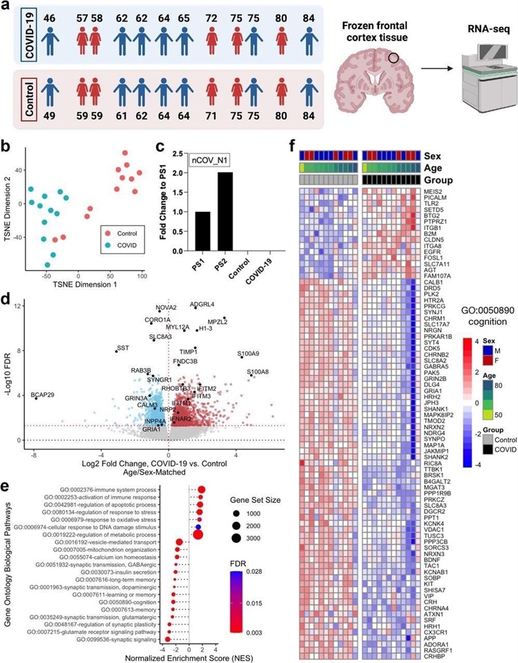
 Severe COVID-19 induces global transcriptomic changes in the frontal cortex of the brain. a. Left, age and sex of each individual in COVID-19 or control groups (n=12/group) analyzed in this cohort. Each COVID-19 case was matched with an uninfected control case by sex and age (±2 years). Right, schematic of the study approach. Schematic was generated with BioRender. b. t-distributed stochastic neighbor embedding (TSNE) analysis of frontal cortex transcriptomes from COVID-19 cases and uninfected controls. c. qPCR assessment of SARS-CoV-2 viral RNA in the frontal cortex using the nCOV_N1 primer set. PS1 and PS2 correspond to the 2019-nCoV_N_Positive Control RUO Plasmid (IDT) at concentrations of 1,000 and 2,000 copies/μl, respectively (a technical duplicate/concentration was used to estimate the corresponding mean; for control and COVID-19 samples n=13/group). d. Volcano plot representing the diffe
rentially expressed genes (DEGs) of the frontal cortex of COVID-19 cases versus matched controls. Red points, significantly upregulated genes among COVID-19 cases (false discovery rate < 0.05). Blue points, significantly downregulated genes among COVID-19 cases. Black points, highlighted significant genes with corresponding gene symbols. e. Gene ontology (GO) biological pathway enrichment analysis of COVID-19 versus control brain DEGs. Gene ranks were determined by signed -log10 false discovery rates of DEGs. FDR, gene set enrichment analysis false discovery rate. f. Heatmap of relative gene expression levels of significant DEGs associated with the “cognition” (GO: 0050890) GO term across COVID-19 and control samples. Color legend, scaled gene expression levels across subjects, normalized via variance-stabilized transformation.
To date, neuroimaging and cognitive screening have identified that COVID-19 induces impairment of the frontal cortex, which plays a vital role in cognitive function. It has also been suggested that COVID-19 results in long-term cognitive impairment. However, the molecular changes induced by COVID-19 that lead to cognitive impairment are yet to be investigated till now.
Typically, natural aging results in decreased frontal cortex activity that may lead to a cognitive deficit. The molecular signatures associated with aging in the human brain are increased immune signaling and decreased synaptic activity.
Interestingly as COVID-19 has been found to cause frontal cortex impairment similar to aging, the study team attempted to investigate if severe COVID-19 resulted in aging-related molecular signatures in the brain.
The study findings showed that severe COVID-19 infection causes distinct global transcriptomic changes in the frontal cortex.
Detailed whole transcriptomic analyses were performed on the postmortem frontal cortex of twelve COVID-19 patients compared with twelve age-matched and sex-matched uninfected controls.
Subsequently clustering analysis via t-distributed stochastic neighbor embedding (TSNE) was performed, which indicated that the transcriptomic profiles of COVID-19 patients were distinct from the controls.
Interestingly, the transcriptomic profiles of two of the controls were aged 71 and 84 were found to be similar to that of the COVID-19 patients.
Detailed quantitative PCR (qPCR) analysis showed the absence of SARS-CoV-2 in the frontal cortex of both the COVID-19 patients and controls at the time of death. This indicates that the gene expression changes observed in COVID-19 patients were not due to the direct action of the virus on the frontal cortex.
The study team identified 2,809 differentially expressed genes (DEGs) bearing unique Ensembl gene IDs when the COVID-19 transcriptomic profiles were compared to age-matched and sex-matched controls. Amongst the DEGs, 1,397 were found to be upregulated, and 1,412 were downregulated.
It should be noted that circulating calprotectin levels is a biomarker that distinguishes severe COVID-19 from mild disease and in the present study, S100A8/S100A9 genes that encode calprotectin were upregulated in COVID-19 patient cohort.
As with earlier reports in COVID-19 patients, SYNGR1 levels were downregulated in the patient cohort. The gene expression changes in COVID-19 patients observed in this study correlate with evidence from previous reports.
Interestingly differentially expressed genes in the frontal cortex of severe COVID-19 patients are similar to those induced by aging
Next, pathway enrichment analysis was performed using annotated Gene Ontology (GO) Biological processes to establish the functional roles of the observed gene expression changes in the transcriptome.
In this analysis, the input gene set is compared with each of the terms or bins in the GO and a statistical test is performed for each term or bin to determine if it is enriched for the input genes.
Importantly the study team observed significant enrichment of several DEGs and GO terms that were related to the aging of the human brain.
In the COVID-19 patient cohort, along with positive enrichment of terms associated with immune response, the related genes such as BCL2, IFI16, and CFH were upregulated.
Also, the expression levels of IFITM1-3, associated with interferon response, were highly dysregulated with elevated levels of expression.
Synaptic function GO terms including synaptic signaling, regulation of synaptic plasticity, glutamatergic, GABAergic, dopaminergic synaptic transmission were found to be negatively enriched, and this correlated with the observed downregulation of genes involved in synaptic signalings such as SST, GRIA1, and GRIN2B.
Significantly, the SST is a gene that has previously been reported to be related to aging in the frontal cortex of humans was observed to be the most downregulated gene in the COVID-19 patient cohort in the present study.
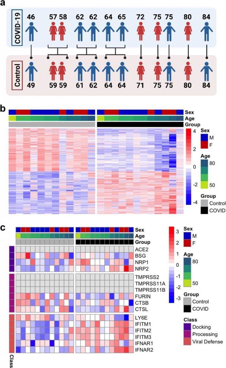
 Overview of differential expression patterns in the frontal cortex of COVID-19 and uninfected subjects. a. Age and sex matching used for differential expression analysis. b. Heatmap of relative gene expression levels of all significant DEGs (FDR < 0.05) across COVID-19 and control samples. Color legend, scaled gene expression levels across subjects, normalized via variance-stabilized transformation. c. Heatmap of relative expression levels of genes previously implicated in SARS-CoV-2 viral entry in the human brain [8] across COVID-19 and control samples. Color legend, scaled gene expression levels across patients, normalized via variance-stabilized transformation. Light gray cells, no aligned reads or genes filtered out of the analysis.
The study findings also showed significant enrichment of GO terms corresponding to the cellular response to DNA damage, mitochondrial function, regulation of response to stress and oxidative stress, vesicular transport, calcium homeostasis, apoptosis, and insulin signaling, and pathways known to be associated with aging and specifically brain aging.
Further, GO terms corresponding to cognitive function, memory, and learning were additionally enriched.
Also the DEGs overlapping with these enriched pathways was assessed, and genes such as the brain-derived neurotrophic factor (BDNF) known to be related to aging were identified.
The Kyoto Encyclopedia of Genes and Genomes (KEGG) and Reactome pathway enrichment analyses were performed, and the findings suggest positive enrichment of immune activation and negative enrichment of synaptic function pathways.
The “Coronavirus Disease-COVID-19” pathway associated with the non-neuronal tissue effects of COVID-19 was identified as a significantly enriched pathway in the COVID-19 patient cohort in the present study.
The study findings suggest that changes in biological pathways that are related to natural aging are also observed in COVID-19 patients with severe disease.
The study findings also revealed that severe COVID-19 induced aging effects are more pronounced in the brains of younger COVID-19 patients.
The study team further investigated if severe COVID-19 induced changes in the transcriptome are similar to the changes induced by aging in the human brain.
The team assessed transcriptome-wide datasets from five patients for aging-related changes and found that genes that were upregulated or downregulated during aging were similarly upregulated or downregulated in COVID-19 patients with severe disease. Particularly a gene set that was known to be associated with aging was significantly upregulated in the COVID-19 patient cohort in this study.
The study findings were further confirmed through quantitative PCR (qPCR) analyses, where genes S100A9, MYL12A, and RHOBTB3 were found to be upregulated and genes CALM3, INPP4A, GRIA1, and GRIN3A were found to be downregulated in the frontal cortex of COVID-19 patients.
Strangely, the same set of genes were also differentially expressed in the frontal cortex from aged individuals.
The study team further attempted to study if the aging gene signature differs between the younger COVID-19 patients aged 65 years or below and the older COVID-19 patients aged above 65 years of age.
In young COVID-19 patients, the study team observed higher levels of changes in gene expression when compared to the older COVID-19 patients.
However, in the case of younger COVID-19 patients, 1,631 upregulated genes and 2,073 downregulated genes were identified that matched with several DEGs of age/sex-matched controls. Whereas in older COVID-19 patients, upregulated expression of 19 genes, including HBA1, HBA2, and HBB genes, and downregulated expression of 4 genes were observed.
Importantly these DEGs also exhibit the same trends associated with aging-related genes in the frontal cortex. These findings indicate that COVID-19 induced aging effects are more pronounced in the brains of younger COVID-19 patients than older ones.
The team suggested that further studies with young COVID-19 patient cohorts will confirm the findings observed.
The study team also investigated if the COVID-19 induced molecular changes differed based on gender.
The team found that the COVID-19 induced changes in aging-related genes and pathways consistent between male and female COVID-19 patients.
This study is the first to demonstrate similarities in the transcriptomic profiles of the frontal cortex of COVID-19 patients and the aging human brain.
The study findings from the present study suggest that severe COVID-19 disease may result in aging-related changes in the brain and may result in premature aging. These changes are more profound in younger patients compared to older ones. Earlier reports indicate a residual cognitive deficit in recovered COVID-19 patients.
The research findings further indicate that increased rates of cognitive impairment and neurodegeneration may occur as long-term effects of long COVID. Therefore, it may be necessary to monitor recovered COVID-19 patients for aging related neurological disorders regularly.
Please help to sustain this site and also all our research and community initiatives by making a donation. Your help means a lot and helps saves lives directly and indirectly and we desperately also need financial help now.
https://www.thailandmedical.news/p/sponsorship
For the latest
COVID-19 Research, keep on logging to Thailand Medical News.

 Severe COVID-19 induces global transcriptomic changes in the frontal cortex of the brain. a. Left, age and sex of each individual in COVID-19 or control groups (n=12/group) analyzed in this cohort. Each COVID-19 case was matched with an uninfected control case by sex and age (±2 years). Right, schematic of the study approach. Schematic was generated with BioRender. b. t-distributed stochastic neighbor embedding (TSNE) analysis of frontal cortex transcriptomes from COVID-19 cases and uninfected controls. c. qPCR assessment of SARS-CoV-2 viral RNA in the frontal cortex using the nCOV_N1 primer set. PS1 and PS2 correspond to the 2019-nCoV_N_Positive Control RUO Plasmid (IDT) at concentrations of 1,000 and 2,000 copies/μl, respectively (a technical duplicate/concentration was used to estimate the corresponding mean; for control and COVID-19 samples n=13/group). d. Volcano plot representing the diffe
rentially expressed genes (DEGs) of the frontal cortex of COVID-19 cases versus matched controls. Red points, significantly upregulated genes among COVID-19 cases (false discovery rate < 0.05). Blue points, significantly downregulated genes among COVID-19 cases. Black points, highlighted significant genes with corresponding gene symbols. e. Gene ontology (GO) biological pathway enrichment analysis of COVID-19 versus control brain DEGs. Gene ranks were determined by signed -log10 false discovery rates of DEGs. FDR, gene set enrichment analysis false discovery rate. f. Heatmap of relative gene expression levels of significant DEGs associated with the “cognition” (GO: 0050890) GO term across COVID-19 and control samples. Color legend, scaled gene expression levels across subjects, normalized via variance-stabilized transformation.
 Overview of differential expression patterns in the frontal cortex of COVID-19 and uninfected subjects. a. Age and sex matching used for differential expression analysis. b. Heatmap of relative gene expression levels of all significant DEGs (FDR < 0.05) across COVID-19 and control samples. Color legend, scaled gene expression levels across subjects, normalized via variance-stabilized transformation. c. Heatmap of relative expression levels of genes previously implicated in SARS-CoV-2 viral entry in the human brain [8] across COVID-19 and control samples. Color legend, scaled gene expression levels across patients, normalized via variance-stabilized transformation. Light gray cells, no aligned reads or genes filtered out of the analysis.