BREAKING! Multisystem Screening Study Finds SARS-CoV-2 Proliferation In Neurons Of The Myenteric Plexus And In Megakaryocytes!
Source: Medical News - SARS-CoV-2 & Myenteric Plexus Apr 03, 2022 3 years, 8 months, 1 week, 5 days, 9 hours, 53 minutes ago
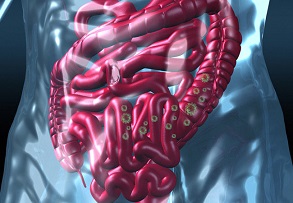
A new study by researchers from Imperial College London, Hammersmith Hospital-London, UK Charing Cross Hospital Campus, UK Chelsea and Westminster NHS Foundation Trust, United Kingdom Health Security Agency, University Hospitals Birmingham and King's College London, has found that the SARS-Cov-2 proliferates in neurons of the myenteric plexus and also in megakaryocytes! This discovery has lots of implications in terms of accounting for extrapulmonary involvement, such as in the gastrointestinal tract and nervous system, as well as frequent thrombotic events in SARS-CoV-3 infected individuals.

Thailand
Medical News would like to add that the discovery of SARS-CoV-2 proliferation in neurons of the myenteric plexus could also be a warning about the virus possibly contributing to the early onset of neurodegenerative diseases like Parkinson’s Disease!
The myenteric plexus (also known as the Auerbach plexus) refers to a network of nerves between the layers of the muscular propria in the gastrointestinal system.
The myenteric plexus provides motor innervation to both layers of the muscular layer of the gut, having both parasympathetic and sympathetic input (although present ganglion cell bodies belong to parasympathetic innervation, fibers from sympathetic innervation also reach the plexus), whereas the submucous plexus has only parasympathetic fibers and provides secretomotor innervation to the mucosa nearest the lumen of the gut.
In simple terms, the myenteric plexus helps regulate peristalsis in the gastrointestinal tract
The myenteric plexus arises from cells in the vagal trigone also known as the nucleus ala cinerea, the parasympathetic nucleus of origin for the tenth cranial nerve (vagus nerve), located in the medulla oblongata. The fibers are carried by both the anterior and posterior vagal nerves. The myenteric plexus is the major nerve supply to the gastrointestinal tract and controls GI tract motility.
A part of the enteric nervous system, the myenteric plexus exists between the longitudinal and circular layers of muscularis externa in the gastrointestinal tract. It is found in the muscles of the esophagus, stomach, and intestine.
The ganglia found in the myenteric plexus have properties similar to the central nervous system (CNS). These properties include presence of glia, interneurons, a small extracellular space, dense synaptic neuropil, isolation from blood vessels, multiple synaptic mechanisms and multiple neurotransmitters.
Megakaryocytes are cells in the bone marrow responsible for making platelets, which are necessary for blood clotting.
The study team performed a comprehensive validation of a monoclonal antibody against the SARS-CoV-2 nucleocapsid protein (NP) followed by systematic multisystem organ immunohistochemistry analysis of the viral cellular tropism in tissue from 36 patients, 16 postmortem cases and 16 biopsies with polymerase chain reaction (PCR)-confirmed SARS-CoV-2 status from the peaks of the pandemic in 2020 and four pre-COVID postmortem controls.
Shockingly, SARS-CoV-2 anti-NP staining in the postmortem cases revealed broad multiorgan involvement of the respiratory, digestive, haematopoietic, genitourinary and nervous systems, with a typical pattern of staining characteriz
ed by punctate paranuclear and apical cytoplasmic labelling.
The average time from symptom onset to time of death was shorter in positively versus negatively stained postmortem cases (mean = 10.3 days versus mean = 20.3 days, p = 0.0416, with no cases showing definitive staining if the interval exceeded 15 days).
A surprising finding was the widespread presence of SARS-CoV-2 NP in neurons of the myenteric plexus, a site of high ACE2 expression, the entry receptor for SARS-CoV-2, and one of the earliest affected cells in Parkinson's disease.
Also, in the in the bone marrow, the study findings showed viral SARS-CoV-2 NP within megakaryocytes, key cells in platelet production and thrombus formation.
In 15 tracheal biopsies performed in patients requiring ventilation, there was a near complete concordance between immunohistochemistry and PCR swab results.
The study findings have relevance to correlating clinical symptoms with the organ tropism of SARS-CoV-2 in contemporary cases as well as providing insights into potential long-term complications of COVID-19.
The study findings were published in the peer reviewed publication: The Journal of Pathology
https://onlinelibrary.wiley.com/doi/full/10.1002/path.5878
It was already previously speculated that, due to the expression of ACE2 in neurons of the myenteric plexus, these cells are possible targets of SARS-CoV-2 infection. The study findings help corroborate this earlier hypothesis.
https://pubmed.ncbi.nlm.nih.gov/33122999/
However, the long-term consequences of this study findings in the myenteric plexus are yet to be established.
Parkinson’s Disease or PD is a neurodegenerative disorder thought to result from the prion-like spread of amyloidogenic alpha-synuclein causing catastrophic motor impairment and other symptoms, such as dementia, as the spread of misfolded protein progresses throughout the brain.
https://pubmed.ncbi.nlm.nih.gov/20493564/
To date however, it is not known if the initial seeding of the misfolded protein conformation is purely a stochastic event or ignited by an internal or external factor, with the exception of 5–10% of cases in which genetic mutations have been shown to impact alpha-synuclein biology.
https://pubmed.ncbi.nlm.nih.gov/16081529/
Importantly, the brain pathology of PD initiates in the olfactory bulb and medulla from where it propagates in a stereotypical pattern, and this is anticipated by accumulation of misfolded alpha-synuclein in the ENS.
This pattern of pathology progression explains anosmia and gastrointestinal (GI) symptoms such as constipation and nausea, appearing early in PD.
https://pubmed.ncbi.nlm.nih.gov/23263478/
It has been proposed that an environmental agent reaches the brain from the nose through the olfactory nerves or from the gut through the ENS via retrograde axonal transport along the vagus nerve reaching the medulla.
https://pubmed.ncbi.nlm.nih.gov/17961138/
https://onlinelibrary.wiley.com/doi/full/10.1002/path.5878
Importantly viral infection of myenteric neurons, as implied by this study findings, could in theory be one such environmental factor.
Correlation between viral infection and Parkinsonism were seen following the 1918 influenza–encephalitis lethargica pandemic.
https://pubmed.ncbi.nlm.nih.gov/18760350/
In this current COVID-19 pandemic, there have been reports of concurrent neurological complications and encephalitis, however, there is currently only one report of postviral Parkinsonism development.
https://pubmed.ncbi.nlm.nih.gov/32637987/
https://pubmed.ncbi.nlm.nih.gov/32949534/
Diversity of genetic background, comorbidities, and previous infections all impact susceptibility and likely may only have neurodegenerative ramifications in a subset of people.
.jpg) Presence of SARS-CoV-2 in neurons of the myenteric plexus and in megakaryocytes in the bone marrow. (A) NP immunostaining of ganglia of the myenteric plexus (PM6). (B) Higher magnification reveals punctate staining within neuronal soma (arrows). Red arrow shows a negative neuron. (C) Overview of extensive positivity within the myenteric plexus. Black arrow points to concentric muscle layer, red arrow points to longitudinal muscle layer. (D) NP staining in neurons of oesophageal myenteric ganglia (PM6). (E) NP staining in megakaryocytes (arrow) with their distinct lobular nuclei. The inset shows the typical paranuclear cytoplasmic punctate pattern (PM10). (F) SARS-CoV-2 in megakaryocytes (see arrow and inset, PM3) is confirmed by colocalisation of NP (brown) with the megakaryocyte marker CD61 (blue). Scale bars = 50 μm.
Presence of SARS-CoV-2 in neurons of the myenteric plexus and in megakaryocytes in the bone marrow. (A) NP immunostaining of ganglia of the myenteric plexus (PM6). (B) Higher magnification reveals punctate staining within neuronal soma (arrows). Red arrow shows a negative neuron. (C) Overview of extensive positivity within the myenteric plexus. Black arrow points to concentric muscle layer, red arrow points to longitudinal muscle layer. (D) NP staining in neurons of oesophageal myenteric ganglia (PM6). (E) NP staining in megakaryocytes (arrow) with their distinct lobular nuclei. The inset shows the typical paranuclear cytoplasmic punctate pattern (PM10). (F) SARS-CoV-2 in megakaryocytes (see arrow and inset, PM3) is confirmed by colocalisation of NP (brown) with the megakaryocyte marker CD61 (blue). Scale bars = 50 μm.
However, as we begin to recover from the pandemic, we should nevertheless still be conscious of COVID-19 when assessing future neurological cases.
Although the extent of SARS-CoV-2's direct invasion of the CNS is uncertain, throughout the pandemic, neurological symptoms such as headache, ageusia, and anosmia were commonly reported, and the involvement of both the central and peripheral nervous system was hypothesized.
https://pubmed.ncbi.nlm.nih.gov/33974053/
In this study, the brain cohort of SARS-CoV-2-infected patients includes patients with clinical and pathological neurological disease, including brainstem encephalitis and cases with no neurological involvement. However, positivity within the brain appeared to be limited to endothelial cells. Crucially, despite observing viral NP in the neurons of the myenteric plexus, the study team found the origin of the vagus nerve in the medulla oblongata to be devoid of neuronal SARS-CoV-2 NP. This suggests that, in the event of any neurodegenerative disease developing after SARS-CoV-2 infection, consideration should be given to the spread of misfolded protein or other pathophysiological alterations triggered by the virus in ENS neurons, rather than the direct entry of the virus into CNS cells.
Past reports about the possibility of viral protein or RNA in the CNS are conflicting.
A certain hypothesis postulates that systemic vascular changes are likely behind reported neurological symptoms, as an indirect consequence of lung-derived hypoxia rather than direct viral damage. Additionally, endothelial cell targeting and injury are also considered to be key elements in COVID-19 pathophysiology that have been frequently alluded to.
https://pubmed.ncbi.nlm.nih.gov/32325026/
The COVID-19 disease has also been established as a prothrombotic disease characterized by coagulopathy, thrombosis, and platelet activation.
https://pubmed.ncbi.nlm.nih.gov/32302448/
https://pubmed.ncbi.nlm.nih.gov/33251499/
The study findings parallel this with findings of platelet-producing megakaryocytes colocalizing with the viral NP within the bone marrow.
Many past studies have also reported the presence of circulating megakaryocytes in multiple organs, including lung, heart, kidney, liver, and brain.
https://pubmed.ncbi.nlm.nih.gov/32895961/
https://pubmed.ncbi.nlm.nih.gov/32766543/
https://pubmed.ncbi.nlm.nih.gov/33576767/
https://pubmed.ncbi.nlm.nih.gov/33307078/
Another study demonstrated that SARS-CoV-2 infection can promote procoagulant changes in platelet gene expression.
https://pubmed.ncbi.nlm.nih.gov/32573711/
Paradoxically, despite confirming viral presence, the study team also ascertained that megakaryocytes and platelets lack ACE2, by which they suggested an independent route of infection. A recent preprint report that infected calprotectin-expressing megakaryocytes additionally express the ACE2 receptor and TMPRSS2.
https://www.medrxiv.org/content/10.1101/2021.08.05.21261552v1
Even more intriguing, that study also provided evidence of megakaryocytes harboring infected pro-platelets. Together with this study findings, it is probable that entry occurs by several mechanisms. The principal molecular route of SARS-CoV-2 entry into megakaryocytes and platelets is therefore still unresolved.
The study findings imply appreciable involvement of the GI tract and related digestive organs in COVID-19, correlating with the high prevalence of symptoms such as diarrhea, abdominal pain, anorexia, and vomiting in COVID-19 symptomatology.
https://pubmed.ncbi.nlm.nih.gov/32405603/
Importantly, the viral cell tropism in the digestive tract overall mirrors that of the expression profile of ACE2 in cell types such as enterocytes.
https://pubmed.ncbi.nlm.nih.gov/15141377/
Preexisting chronic liver diseases, such as steatohepatitis and cirrhosis, have been commonly associated with severe cases of COVID-19, with 2–11% of patients having liver comorbidities.
https://pubmed.ncbi.nlm.nih.gov/32145190/
https://onlinelibrary.wiley.com/doi/full/10.1002/path.5878
With four of ten patients displaying liver disease, this is also a common finding within this full-body postmortem cohort. In addition to endothelial cells, three cases (PM1, PM6, and PM10) exhibited a striking viral NP presence within parenchymal tissues, including bile duct cholangiocytes, as well as hepatocytes.
Studies of the ACE2 receptor in normal liver asserts low expression in hepatocytes; however, mouse models and human samples suggest chronic liver injury may contribute to ACE2 receptor upregulation.
https://pubmed.ncbi.nlm.nih.gov/15141377/
https://pubmed.ncbi.nlm.nih.gov/16166274/
Cholangiocytes, on the other hand, purportedly have high levels of ACE2, which may explain the strong viral protein detection seen in this study.
The study team concluded, “Most significantly, our study findings provide evidence of neuro-invasiveness in the ENS but not the CNS. We have also identified megakaryocytes as potential targets of SARS-CoV-2. Further investigations in gene expression profiling related to these findings will help determine any long-term implications.”
For more the latest
SARS-CoV-2 research, keep on logging to Thailand medical News.

.jpg) Presence of SARS-CoV-2 in neurons of the myenteric plexus and in megakaryocytes in the bone marrow. (A) NP immunostaining of ganglia of the myenteric plexus (PM6). (B) Higher magnification reveals punctate staining within neuronal soma (arrows). Red arrow shows a negative neuron. (C) Overview of extensive positivity within the myenteric plexus. Black arrow points to concentric muscle layer, red arrow points to longitudinal muscle layer. (D) NP staining in neurons of oesophageal myenteric ganglia (PM6). (E) NP staining in megakaryocytes (arrow) with their distinct lobular nuclei. The inset shows the typical paranuclear cytoplasmic punctate pattern (PM10). (F) SARS-CoV-2 in megakaryocytes (see arrow and inset, PM3) is confirmed by colocalisation of NP (brown) with the megakaryocyte marker CD61 (blue). Scale bars = 50 μm.
Presence of SARS-CoV-2 in neurons of the myenteric plexus and in megakaryocytes in the bone marrow. (A) NP immunostaining of ganglia of the myenteric plexus (PM6). (B) Higher magnification reveals punctate staining within neuronal soma (arrows). Red arrow shows a negative neuron. (C) Overview of extensive positivity within the myenteric plexus. Black arrow points to concentric muscle layer, red arrow points to longitudinal muscle layer. (D) NP staining in neurons of oesophageal myenteric ganglia (PM6). (E) NP staining in megakaryocytes (arrow) with their distinct lobular nuclei. The inset shows the typical paranuclear cytoplasmic punctate pattern (PM10). (F) SARS-CoV-2 in megakaryocytes (see arrow and inset, PM3) is confirmed by colocalisation of NP (brown) with the megakaryocyte marker CD61 (blue). Scale bars = 50 μm.