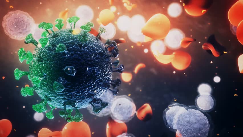
BREAKING NEWS! COVID-19 Study Reveals That SARS-CoV-2 Uses CD4 Cells To Infect T-helper Lymphocytes. COVID-19 A Potent Version Of Airborne HIV?
Source: BREAKING NEWS Sep 30, 2020 5 years, 3 months, 1 week, 13 hours, 58 minutes ago
Breaking News: A multi-disciplinary team of researchers from University of Campinas (UNICAMP)- Brazil have in a new study discovered and proved that the SARS-CoV-2 infects human CD4+ T helper cells, but not CD8+ T cells, and is present in blood and bronchoalveolar lavage T helper cells of severe COVID-19 patients.

The study findings are published on a preprint server but are already being peer-reviewed for publication in a leading journal.
https://www.medrxiv.org/content/10.1101/2020.09.25.20200329v1#disqus_thread
Thailand Medical news first gave a heads up that the SARS-CoV-2 coronavirus was able to infect the CD4 cells as early as April 16
th based on a research by Chinese researchers that was published in Nature journal. However that research was retracted by the authors themselves on basis of the T Cells lines used and they strangely never wanted to pursue the study again using primary T Cells. They further made claims themselves that there were flaws in their flow cytometry methodology.
https://www.thailandmedical.news/news/covid-19-alert-new-study-shows-sars-cov-2-coronavirus-targets-and-destroys-t-cells,-similarly-as-what-hiv-does
Retraction-
https://www.nature.com/articles/s41423-020-0424-9
Also strangely all studies concerning the effects of the new SARS-CoV-2 virus on T Cells and CD4 cells were removed from public domains and online research journals in China then, despite the fact the Pasteur Institute Of Virology In Shanghai and the Chinese Academy of Sciences was doing extensive studies on that area still. It interesting as to what the Chinese authorities really know that the rest of the world does not with regards to the SARS-CoV-2 coronavirus.
Another coverage based on a study by Britains Kings College was also downplayed and disregarded by numerous health agencies and research entities.
https://www.thailandmedical.news/news/covid-19-research-new-study-reveals-that-specific-types-of-t-cells-are-targeted-by-the-sars-cov-2-coronavirus-in-patient-s-immune-system
Last week Dr Anthony Fauci at a senator hearing started to distance himself on an earlier hypothesis that he was touting in the past with regards to T cell protective immunity.
https://www.thailandmedical.news/news/covid-19-headlines-why-is-dr-fauci-now-distancing-himself-from-an-earlier-t-cell-immunity-hypothesis-he-was-previously-peddling
In reality it seems that certain world leaders and authorities, and even huge pharma companies are aware of the magnitude of the COVID-19 disease but are simply downplaying the issues and simply buying time. Even in terms of genomic studies showing the emerging n
ew strains and the mutations taking place with possible implications are all being ignored despite scientifc evidence by credible researchers and institutions.
In the new Brazilian study that involved over 68 scientists willing to put their reputations at stake, it was demonstrated that SARS-CoV-2 infects human CD4+ T helper cells, but not CD8+ T cells, and is present in blood and bronchoalveolar lavage T helper cells of severe COVID-19 patients.
The team demonstrated that SARS-CoV-2 spike glycoprotein (S) directly binds to the CD4 molecule, which in turn mediates the entry of SARS- CoV-2 in T helper cells in a mechanism that also requires ACE2 and TMPRSS2. Once inside T helper cells, SARS-CoV-2 assembles viral factories, impairs cell function and may cause cell death.
Interestingly SARS-CoV-2 infected T helper cells express higher amounts of IL-10, which is associated with viral persistence and disease severity. Thus, CD4-mediated SARS-CoV-2 infection of T helper cells may explain the poor adaptive immune response of many COVID- 19 patients.
Individuals that progress to the severe stages of COVID-19 that is marked alterations in the immune response characterized by reduced overall protein synthesis, cytokine storm, lymphocytopenia and T cell exhaustion. Besides these acute effects on the immune system, most convalescent individuals present low titers of neutralizing antibodies. Also, the levels of antibodies against SARS-CoV-2 decay rapidly after recovery, suggesting that SARS-CoV-2 infection may exert profound and long-lasting complications to adaptive immunity. In this context, one question that remains to be answered is how SARS-CoV-2 exerts these effects on the immune system.
In order to infect cells, the spike glycoprotein of SARS-CoV-2 binds to the host angiotensinconverting enzyme 2 orACE2, after which it is then cleaved by TMPRSS2 11. While TMPRSS2 is ubiquitously expressed in human tissues, ACE2 is mainly expressed in epithelial and endothelial cells, as well as in the kidney, testis and small intestine. Even then a wide variety of cell types are infected by SARS-CoV-212-14, even though some of these cells express very low levels of ACE2. The study team showed that this is the case for lymphocytes.
This finding suggests that SARS-CoV-2 has either an alternative mechanism to enter the cells or that auxiliary molecules at the plasma membrane may fix the virus until it interacts with an ACE2 molecule. Since the structures of the spike of SARS-CoV-1 and the SARS-Cov-2 proteins are similar, the team used the P-HIPSTer algorithm to uncover human proteins that putatively interact with the viruses.
Seventy-one human proteins were predicted to interact with SARS-CoV-1. The team then cross-referenced the proteins with five databases of plasma membrane proteins to identify the ones located on the cell surface.
CD4 was the only protein predicted to interact with SARS-CoV-1that appeared in all five databases. CD4 is expressed mainly in T helper lymphocytes and has been shown to be the gateway for HIV.
Since CD4+ T lymphocytes orchestrate innate and adaptive immune responses infection of CD4+ T cells by SARS-CoV-2 might explain lymphocytopenia and dysregulated inflammatory response in severe COVID-19 patients.
From an evolutionary perspective, the infection of CD4+ T cells represents an effective mechanism for viruses to escape the immune response. To test whether human primary T cells are infected by SARS-CoV-2, the team purified CD3+ CD4+ and CD3+ CD8+ T cells from the peripheral blood of non-infected healthy controls/donors (HC), incubated these cells with SARS-CoV-2 for 1h, and then exhaustively washed them to remove any residual virus. The viral load was measured 24h post-infection. The team was able to detect SARS-CoV-2 RNA in primary CD4+ T cells but not CD8+ T cells.
To confirm the presence of SARS-CoV-2 infection, the team performed in situ hybridization using probes against the viral RNA-dependent RNA polymerase (RdRp) gene, immunofluorescence for sCoV-2 and transmission electron microscopy. All three approaches confirmed that SARS-CoV-2 infects CD4+ T cells.
The team also detected different SARS-CoV-2 RNAs in infected CD4+ T cells. Notably, the viral RNA level increases with time and they identified the presence of the negative strand (antisense) of SARS-CoV-2 in the infected cell, demonstrating that the virus is able to assemble viral factories and replicate in T helper cells.
Plaque assay also revealed that SARS-CoV-2-infected CD4+ T cells produce infectious viral particles. To confirm that SARS-CoV-2 infects CD4+ T cells in vivo, the team purified CD4+ and CD8+ T cells from peripheral blood cells of COVID-19 patients.
Similar to the ex a vivo finding, SARS-CoV-2 RNA was detected in CD4+ T cells, but not in CD8+ T cells from COVID-19 patients. Yet, the viral load was markedly higher in CD4+ T cells from severe COVID19 patients in comparison to patients with the moderate form of the disease.
Utilizing publicly available single-cell sequencing data22, the study team also able to detect the presence of SARS-CoV-2 RNA in 2.1% of CD4+ T cells of the bronchoalveolar lavage (BAL) of patients with the severe but not the moderate form of COVID-19.
Hence the study data demonstrates that SARS-CoV-2 infects CD4+ T cells and the infection associates with the severity of COVID19.
The team sought to explore the role of the CD4 molecule in SARS-CoV-2 infection. Based on the putative interaction found using P-HIPSTer, they performed molecular docking analyses and predicted that sCoV-2 receptor binding domain (RBD) directly interacts with the N-terminal domain (NTD) of CD4 Ig-like V type. Molecular dynamics simulations with stepwise temperature increase were applied to challenge the kinetic stability of the docking model representatives. Two models remained stable after the third step of simulation at 353 Kelvin and represent likely candidates for the interaction between the CD4 NTD and sCoV-2 RBD. Additionally, convergence towards the two surviving models was tested for closely related binding mode models present among the remaining cluster candidates and was verified in one case, which indicates plausible and rather stable interaction between CD4 NTD and sCoV-2 RBD .
The interaction region of RBD is predicted to overlap with that of human ACE2. The interaction between CD4 and sCoV-2 was confirmed by co-immunoprecipitation of sCoV-2 and full length recombinant CD4.
Consistent with a mechanism where CD4-sCoV-2 interaction is required for infection, the team observed that increasing concentrations of soluble CD4 (sCD4) reduced CD4+ T cell infection by SARS-CoV-2. To gain further insights into the importance of CD4-sCoV-2 binding to SARS-CoV-2 infection, the team purified CD4+ T cells and pre-treated them with anti-CD4 polyclonal antibody.
The team observed a dose-dependent reduction in viral load in CD4+ T cells pre-treated with anti-CD4 antibody, showing that CD4 is necessary for SARS-CoV-2 infection.
Remarkably, the same monoclonal antibody that has been used to block HIV entry in CD4+ T cells also blocked SARS-CoV-2 entry in a dose dependent manner.
This observation is consistent with our in silico model that predicts that sCoV-2 binds to a region of CD4 which is neighbor to where envelope-displayed glycoprotein spike complex (Env) is shown to bind 24.
These data demonstrate that the CD4 molecule is necessary for infection of CD4+ T cells by SARS-CoV-2 and suggest that
SARS-CoV-2 may use a mechanism that somehow resembles HIV infection.
In HIV infection, CD4 alone is not sufficient to allow the virus to enter CD4+ T cells25. Instead, the Env must also interact with co-receptors (CCR5 or CXCR4).
In this context, the team tested whether CD4 alone was sufficient to allow SARS-CoV-2 entry. Inhibition of ACE2 using polyclonal antibody abrogated SARS-CoV-2 entry in CD4+ T cells, suggesting that the canonical entry mechanism involving ACE2 and TMPRSS211 is also required. To exclude the possibility that the polyclonal anti-ACE2 antibody cross-reacts with CD4, the team designed a peptide to specifically block ACE2-sCoV-2 interaction .
That peptide recapitulated the effect of anti-ACE2 antibody and also reduced the viral load in a dose dependent manner.
Similarly, inhibition of TMPRSS2 with camostat mesylate also reduced SARS-CoV-2 infection. Hence, ACE2, TMPRSS2 and CD4 act in concert to allow the infection of CD4+ T cells by SARS-CoV-2.
To assess the consequences of SARS-CoV-2 infecting CD4+ T cells, the team performed mass spectrometry-based shotgun proteomics in ex vivo infected CD4+ T cells.
They found that SARS-CoV-2 infection affects multiple housekeeping pathways associated with the immune system, infectious diseases, cell cycle and cellular metabolism, similarly to what was observed in HIV-infected CD4+ T cells.
SARS-CoV-2 infection elicits alterations associated with “cellular responses to stress”, which include changes in proteins involved in translation, mitochondrial metabolism, cytoskeleton remodeling, cellular senescence and apoptosis. Consistent with these changes, ex vivo infection of CD4+ T cells resulted in a decrease of 10% in cell viability 24h after infection even with a low MOI (0.1). The infection of CD4+ T cells by HIV also causes an increase in IL-10 production. The expression and release of IL-10 has been widely associated with chronic viral infections and determines viral persistence.
Noteworthy, increased serum levels of IL-10 are associated with COVID-19 severity. The team found that IL10 expression by CD4+ T cells was higher in BAL and blood of severe COVID-19 patients. These changes were at least in part cell autonomous, since purified CD4+ T cells infected ex vivo with SARS-CoV-2 also expressed higher levels of IL10 .
Due to the immunomodulatory role of IL-10, the team measured the expression of key pro- and anti-inflammatory cytokines involved in the immune response elicited by CD4+ T cells. CD4+ T cells from severe COVID-19 patients presented decreased expression of IFNG and IL17A in relation to cells from patients with the moderate form of the disease or healthy donors .
These results show that SARS-CoV-2 infection induces IL10 expression in CD4+ T cells. This phenomenon is associated with a suppression of genes that encode key pro-inflammatory cytokines produced by CD4+ T cells, such as IFNγ and IL-17A, and correlates with disease severity.
Upregulation of IL10 in HIV-infected cells requires STAT3-independent activation of the transcription factor CREB-1 via Ser133 phosphorylation33.
Consistent with the similarities between SARS-CoV-2 and HIV infections, CREB-1 phosphorylation at Ser133 was increased in SARS-CoV-2-infected CD4+ T cells.
Hence SARS-CoV-2 infection appears to directly trigger a signaling cascade that culminates in upregulation of IL10 in CD4+ T cells. Indeed, expression of IL10 is positively correlated with viral load in circulating CD4+ T cells from COVID-19 patients.
Altogether, the study data demonstrate that SARS-CoV-2 infects CD4+ T cells, impairs cell function, leads to increased IL10 expression and compromises cell viability, which in turn dampens immunity against the virus and contributes to disease severity. Impaired innate and adaptive immunity is a hallmark of COVID-19, particularly in patients who progress to the critical stages of the disease. Here we show that the alterations in immune response associated with the severe illness from COVID-19 are triggered by infection of CD4+ T helper cells by SARS-CoV-2 and consequent dysregulation of immune function. T helper cells are infected by SARS-CoV-2 using a mechanism that involves binding of sCoV2 to CD4 and entry via the canonical ACE2/TMPRSS2 pathway.
The study model suggests the hypothesis that CD4 stabilizes SARS-CoV-2 on the cell membrane until the virus encounters an ACE2 molecule to enter the cell. This mechanism is similar to what has been described for HIV infection.
Once in CD4+ T cells, SARS-CoV-2 leads to protein expression changes consistent with alterations in pathways related to stress response, apoptosis and cell cycle regulation which, if sustained, culminate in cell dysfunction and may lead to cell death.
SARS-CoV-2 infection also results in phosphorylation of CREB-1 transcription factor and induction of its target gene IL10 in a cell autonomous manner. Yet again, this mechanism resembles HIV infection.
IL-10 is a powerful anti-inflammatory cytokine and has been previously associated with viral persistence. Serum levels of IL-10 increase during the early stages of the disease – when viral load reaches its peak – and may predict COVID-19 outcome.
This increase occurs only in patients with the severe form of COVID-19. Consistent with these findings, the team found that expression of IL10 positively correlates with viral load in CD4+ T cells.
This is a unique feature of patients with the severe form of COVID-19, since the team could not detect the virus in CD4+ T cells from patients with the moderate form of the disease and IL10 expression in CD4+ T cells is much lower in these patients.
In contrast, the team found IFNG and IL17A to be upregulated in CD4+ T cells of patients with the moderate illness, indicating a protective role for these cytokines.
However, in patients with the severe symptoms, the expression of IFNG and IL17A in CD4+ T cells is dampened. IL-10 is a known suppressor of Th1 and Th17 responses and it is likely to contribute to the changes in IFNG and IL17A.
These features will ultimately reflect in the quality of the immune response, which in combination with T cell death and consequent lymphopenia, may result in transient/acute immunodeficiency and impair adaptive immunity in severe COVID-19 patients.
How long these alterations in T cell function persist in vivo and whether they have long-lasting impacts on adaptive immunity remains to be determined.
Hence, avoiding T cell infection by blocking sCoV-2-CD4 interaction and boosting T cell resistance against SARS-CoV-2 might represent complementary therapeutic approaches to preserve immune response integrity and prevent patients from progressing to the severe stages of COVID-19.
This new research finding has massive implications and changes everything on how the disease should be treated to even the effects that the virus has on the long term health implications of ‘recovered’ COVID-19 patients.
For more
BREAKING NEWS on the COVID-19 disease, keep on logging to Thailand Medical News.
