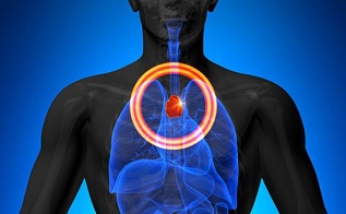
BREAKING! SARS-CoV-2 Causes Thymic Dysregulation And Thymic Atrophy That Results In Lymphopenia And Peripheral T Cell Receptor Repertoire Changes!
Source: Thailand Medical - SARS-CoV-2-T-Cells May 09, 2022 2 years, 11 months, 1 week, 4 days, 21 hours, 2 minutes ago
A new study by Indian researchers from the Translational Health Science and Technology Institute-India, Reginal Centre Biotechnology -India and the All India Institute of Medical Sciences has found that the SARS-CoV-2 coronavirus causes thymic dysregulation and thymic atrophy that results in lymphopenia and peripheral T cell receptor repertoire changes!

It has already been known that most pathogenic infections cause thymic atrophy, perturb thymic-T cell development and alter immunological response.
Numerous past studies have reported dysregulated T cell function and lymphopenia in COVID-19 patients. However, immune-pathological changes, in the thymus, post severe acute respiratory syndrome coronavirus-2 (SARS-CoV-2) infection have not been elucidated.
The study findings alarmingly show that SARS-CoV-2 infects thymocytes, depletes CD4 + CD8+ (double positive; DP) T cell population associated with an increased apoptosis of thymocytes, which leads to severe thymic atrophy in K18-hACE2-Tg mice.
Importantly CD44 + CD25- T cells were found to be enriched in infected thymus, indicating an early arrest in the T cell developmental pathway.
It was also found that Interferon gamma (IFN-γ) was crucial for thymic atrophy, as anti-IFN-g antibody neutralization rescued the loss of thymic involution.
The study also found that the Omicron BA.1 variant of SARS-CoV2 caused marginal thymic atrophy, while the delta variant of SARS-CoV-2 exhibited most profound thymic atrophy characterized by severely depleted DP T cells.
However, the study does not cover the BA.2 variant and numerous emerging subvariants neither does it cover the new BA.4 and BA.5 variant and also many new subvariants of these two new variants.
The study findings provide the first report of SARS-CoV-2 associated thymic atrophy resulting from impaired T cell developmental pathway and also explains dysregulated T cell function in COVID-19.
The study findings were published on a preprint server: Research Square and is currently being peer reviewed.
https://www.researchsquare.com/article/rs-1581769/v1
The study team had wanted to explored the underlying mechanism resulting in thymic atrophy and subsequent lymphopenia in COVID-19 patients.
Numerous past studies have reported dysregulated T cell function and lymphopenia in COVID-19 patients. However, none sheds any light on immunological and pathological alterations in thymus post-severe acute respiratory syndrome coronavirus-2 (SARS-CoV-2) infection.
Corresponding author, Dr Amit Awasthi from the Translational Health Science and Technology Institute-India told
Thailand Medical News, “It should be noted that the thymus is the primary site of T cell development; aging, pathogenic infections, nutritional deficiencies, cancer, and hormonal changes impact its health, which is defined by its output.”
He further added, “Importantly, the peripheral escape of thymocytes, a
rrest in the developmental pathway, or increased apoptosis are all responsible for thymic atrophy due to pathogenic infection.”
For the research, the study team intranasally infected hamsters or human angiotensin-converting enzyme 2 (hACE2)-Tg transgenic mice with 105 PFU of live SARS-CoV-2. The viral inoculum for hamsters and mice was 100 μl for hamsters and 50 μl for hACE2-Tg mice. The control group received Dulbecco's Modified Eagle Medium (DMEM) instead of the live virus, and unchallenged animals received phosphate buffer saline (PBS) injections.
During the study, from a day before viral challenge till day three post-infection, test group animals received a subcutaneous (sc) injection of remdesivir (RDV) at 25 mg/kg body, and control group animals received PBS injections.
The study team used an anti-mouse interferon-gamma (IFNγ) antibody for IFN-γ neutralization at a dosage of 10 mg/kg body mass at two-time points a day before the SARS-CoV-2 challenge and two days post-challenge through intra-peritoneal injections. The control group received immunoglobulin G antibody only.
The study team noted the body mass of the animals daily post-challenge. Likewise, they sacrificed six animals from both test and control groups, and their thymus was extracted and examined for any gross morphological changes.
The study team also analyzed thymus sections using immunofluorescence microscopy for nucleocapsid (N) protein. A trained pathologist scored hematoxylin and eosin (H&E) stained sections on a scale of 0-5, where a score of 5 indicated the highest pathological feature.
The team also performed immunophenotyping of human peripheral blood mononuclear cells (PBMCs).
The researchers also isolated ribonucleic acid (RNA) from homogenized thymus cells, reverse-transcribed these to complementary deoxyribonucleic acid (cDNA), and performed a quantitative polymerase chain reaction (qPCR). They used the cloned transcript (as a template) for generating a standard curve to estimate the copy number of SARS-CoV-2 N gene RNA. The researchers used the trypan-blue exclusion method to determine the number of live cells from the thymus and lymph node.
The study team compared and analyzed thymus and body mass, gene expression, and enzyme-linked immunosorbent assay (ELISA) results using one-way ANOVA or two-way ANOVA.
Interestingly, it was found that the hACE2-Tg mice infected with Wuhan-Hu 1 SARS-CoV-2 strain developed profound thymic atrophy with seven-to-eight folds reduced size, primarily arrested the developing thymocytes in the double-negative 1 (DN1) stage.
Intriguingly, SARS-CoV-2-induced thymocyte apoptosis led to increased cell death.
Alarmingly, the qPCR data showed the presence of SARS-CoV-2 N gene RNA in all the cell subpopulations of thymocytes. Immunofluorescence microscopy also detected the presence of virus in the cellular compartments of thymocytes. However, the investigation could not identify the exact mechanism of SARS-CoV-2 entry into the thymocytes.
The study team hence speculated that either infected progenitor T cells migrated to the thymus, thus disseminating infection, or whether the virus migrated to the thymus through direct impounding.
IFN-γ adequately induced thymic atrophy as neutralization of IFN-γ by neutralizing monoclonal antibodies salvaged thymic atrophy. Whereas, interleukin 17 (IL-17), IL-4, granzyme B (GzB), and perforin-1 (Prf-1) had a limited role in thymic atrophy induced by SARS-CoV-2. Interestingly, antiviral RDV therapy effectively rescued mice from SARS-CoV-2-related thymic atrophy.
Compared to the Omicron BA.1 variant, the Delta-induced severe thymic atrophy was far worse than the ancestral strain in Delta-infected mice, with profoundly impaired T cell development.
The study findings demonstrated that thymic dysregulation and thymic atrophy cause SARS-CoV-2-related lymphopenia and peripheral T cell receptor (TCR) repertoire changes. Since thymic atrophy results in loss of peripheral TCR repertoire, the study findings might improve understanding of how T cell response was reduced during COVID-19 and help develop novel vaccine candidates.
For more on T-Cells and SARS-CoV-2, keep on logging to
Thailand Medical News.
Read Also:
https://www.thailandmedical.news/news/why-is-no-one-warning-the-masses-that-the-sars-cov-2-spike-proteins-are-causing-major-immunodeficiency-issues-in-all-infected-individuals
