BREAKING! University Of Queensland Study Reveals That SARS-CoV-2 Spike Proteins Induces NLRP3 Inflammasome Activation In Human Microglia!
Source: NeuroCOVID - SARS-CoV-2 - Microglia Nov 08, 2022 2 years, 5 months, 1 week, 5 days, 10 hours, 18 minutes ago
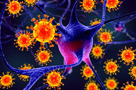
NeuroCOVID: A new study by researchers from the University Of Queensland- Australia has revealed that the spike proteins of the SARS-CoV-2 coronavirus induces NLRP3 inflammasome activation in human microglia!

The study findings have enormous implications and bearings on the long term
NeuroCOVID complications in individuals exposed to the novel coronavirus and also potential increase in current known neurodegenerative diseases and possible the emergence of new ones!
The microglia are the immune cells of the central nervous system and consequently play important roles in brain infections and inflammation and also in neurodegenerative issues.
Although COVID-19 disease is primarily a respiratory disease, an increasing number of reports indicate that SARS-CoV-2 infection can also cause severe neurological manifestations, including precipitating cases of probable Parkinson’s disease.
It is already known that microglial NLRP3 inflammasome activation is a major driver of neurodegeneration and the study team decided to investigate whether SARS-CoV-2 can promote microglial NLRP3 inflammasome activation.
Utilizing SARS-CoV-2 infection of transgenic mice expressing human angiotensin-converting enzyme 2 (hACE2) as a COVID-19 pre-clinical model, the study team established the presence of virus in the brain together with microglial activation and NLRP3 inflammasome upregulation in comparison to uninfected mice.
Subsequently, using a model of human monocyte-derived microglia, the study team surprisingly identified that SARS-CoV-2 isolates can bind and enter human microglia in the absence of viral replication.
Importantly, this interaction of virus and microglia directly induced robust inflammasome activation, even in the absence of another priming signal.
The study team mechanistically demonstrated that purified SARS-CoV-2 spike glycoprotein activated the NLRP3 inflammasome in LPS-primed microglia, in a ACE2-dependent manner.
The spike protein also could prime the inflammasome in microglia through NF-κB signaling, allowing for activation through either ATP, nigericin or α-synuclein.
Significantly, SARS-CoV-2 and spike protein-mediated microglial inflammasome activation was significantly enhanced in the presence of α-synuclein fibrils and was entirely ablated by NLRP3-inhibition.
The study team also demonstrated SARS-CoV-2 infected hACE2 mice treated orally post-infection with the NLRP3 inhibitory drug MCC950, have significantly reduced microglial inflammasome activation, and increased survival in comparison with untreated SARS-CoV-2 infected mice.
The study findings support a possible mechanism of microglial innate immune activation by SARS-CoV-2, which could explain the increased vulnerability to developing neurological symptoms akin to Parkinson’s disease in COVID-19 infected individuals, and a potential therapeutic avenue for intervention.
The study findings were published in the peer revie
wed journal: Molecular Psychiatry.
https://www.nature.com/articles/s41380-022-01831-0
This is among the first few research works to assess the role of severe acute respiratory syndrome coronavirus 2 (SARS-CoV-2) in the NLR family pyrin domain containing 3 (NLRP3) inflammasome activation.
It should be noted that peripheral diseases include Guillain-Barre syndrome, muscle injury resembling myositis, and, most significantly, almost 65% of SARS-CoV-2-infected patients reported hyposmia, which is a common pre-motor symptom in Parkinson's disease (PD).
Furthermore, studies are now determining the association between SARS-CoV-2 infections and Parkinson's disease (PD) in light of documented cases of PD associated with coronavirus disease 2019 (COVID-19).
To date however, the mechanism via which SARS-CoV-2 could raise the risk of manifestation of PD and other neurological problems, as well as how COVID-19 might affect synucleinopathy, is not yet identified.
The study team employed a human monocyte-derived microglia (MDMi) cellular model to examine inflammasome activation observed against SARS-CoV-2 and its spike protein and the expression of microglial NLRP3 inflammasome in the brain following SARS-CoV-2 infection.
The researchers infected female K18 human angiotensin-converting enzyme 2 (hACE2) mice with an early clinical isolate of the SARS-CoV-2 Wuhan strain to assess the virus' impact on the brain. Up to 12 days post-infection (dpi), body weights, clinical scores, and survival rates were documented.
Subsequently, a nanobody that detected divergent SARS-CoV-2 variants, such as the Wu strain, was employed to examine the brains of infected mice with six dpi.
The study team then estimated NLRP3 expression by immunofluorescence to ascertain whether inflammasome activation occurred after SARS-CoV-2 infection.
The detailed contribution of SARS-CoV-2 to the promotion of inflammasome activation within the human microglia was also examined. To obtain adult microglia, MDMi was produced.
The study team confirmed that, compared to monocyte-derived macrophages, the developed MDMi strongly expressed the common microglia hallmark markers, including P2RY12 and TMEM119.
The release of infectious viral particles following infection was estimated using multiplicity of infection (MOI) values of 1 and 0.1 to assess whether these MDMi supported SARS-CoV-2 replication.
The study team exposed the MDMi cells to SARS-CoV-2 and important inflammasome activation signals were estimated to investigate the activation of microglial inflammasomes after SARS-CoV-2 infection.
The MOI of 1 Wuhan isolate was incubated with MDMi or lipopolysaccharide (LPS)-primed cells. Immunocytochemistry and Western blot were used to identify signs of inflammasome activation 24 hours after infection.
The study team next tested whether spike protein could directly activate inflammasomes in human microglia.
The study team created a low endotoxin S-clamp and a control trimeric fusion protein (F-clamp) derived from the Nipah virus. These proteins were verified using sodium dodecyl-sulfate polyacrylamide gel electrophoresis (SDS-PAGE), size exclusion chromatography, and enzyme-linked immunosorbent assay (ELISA) to further understand the mechanism of inflammasome activation by SARS-CoV-2.
The study team detected widespread viral spread in the parenchymal brain cells of infected mice.
Furthermore, ionized calcium-binding adaptor molecule 1 (Iba-1) staining for the pan-microglia/macrophage protein also showed morphological changes indicating microglial activation in brains infected with SARS-CoV-2. When stained for the particular microglial marker TMEM119, SARS-CoV-2-infected brains revealed retracted and thickened TMEM119-positive cell processes along with large cell bodies, indicative of microglial activation. Also, co-staining with anti-SARS-CoV-2 nucleocapsid (N) protein showed that the virus was present in the vicinity of these activated microglia.
The use of qPCR further verified the participation of inflammasomes in SARS-CoV-2-infected brains, with significant overexpression of caspase-1, Aif1 (Iba1), and pycard (ASC).
The study findings demonstrated that SARS-CoV-2 infection in mice activated the microglia and upregulated the inflammasome components of NLRP3.
The inflammasome activation was induced by SARS-CoV-2 infection in MDMi, as evidenced by cleaved interleukin (IL)-1 release in the cells' supernatant after exposure to the Wuhan strain.
This hence supported the inflammasome's activation by correlating with rising levels of cleaved caspase-1. ASC speck formation, a biological indicator of inflammasome activation, supported these findings.
Both Wuhan-treated MDMi and nigericin (Nig)-activated LPS-primed cells used as positive control showed increased ASC speck staining. Notably, SARS-CoV-2 exposure could instantly activate the inflammasome in MDMi without priming, indicating that the virus can prime and activate the inflammasome.
Utilizing size-exclusion chromatography, the study team confirmed that most of the S-clamp and F-clamp proteins were present in their trimeric form and maintained reactivity as measured by the binding of crucial LPS-induced antibodies. These proteins had disintegrated after the LPS wash-out but had been re-induced by the viral spike protein.
Importantly, this showed that the spike protein primed and activated the inflammasome.
The study team also noted that compared to a control protein such as NCAM-FcM, which was generated similarly to hACE2-FcM, the human ACE2 protein prevented SARS-CoV-2 entry with a 50% inhibitory concentration (IC50) of 39 g/mL into the Vero E6 cells.
However, in contrast to CO5 and nigericin, pre-treating LPS-primed MDMi with 3E8 reduced IL-1-beta production following activation with S-clamp. This indicated that spike-ACE2 interaction particularly contributed to inflammasome activation in microglia.
More detailed research is warranted, but this is potentially a new approach to treating a virus that could otherwise have untold long-term health ramifications especially in the area of neurodegenerative and CNS issues and complications.
The study findings established that SARS-CoV-2 and its spike protein could prime and activate the NLRP3 inflammasome present in human microglia. This highlighted the potential COVID-19 risk factor in manifesting neurodegeneration and Parkinson’s Disease.
For the latest on
NeuroCOVID, keep on logging to Thailand
Medical News.
