Columbia Study Reveals That Ferroptosis Plays A Key Role In COVID-19 Pulmonary Disease
Nikhil Prasad Fact checked by:Thailand Medical News Team May 22, 2024 11 months, 4 days, 1 hour, 39 minutes ago
COVID-19 News: As the COVID-19 pandemic continues to impact millions worldwide, scientists race to uncover the underlying mechanisms that lead to severe respiratory complications. A groundbreaking study by Columbia University that is covered in this
COVID-19 News report has revealed a critical role played by ferroptosis, an iron-dependent form of cell death, in the pulmonary manifestations of COVID-19. This discovery opens new avenues for potential treatments aimed at mitigating lung damage caused by SARS-CoV-2.
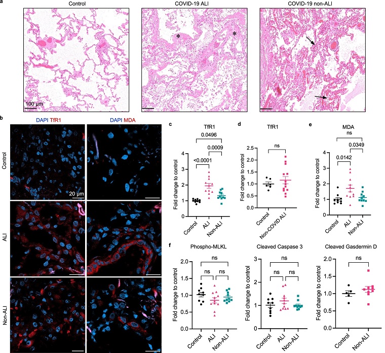
 Fatal COVID-19 pulmonary disease involves ferroptosis
a Representative images of H&E-stained COVID-19 lung autopsies with ALI and non-ALI pathology and non-infected control lungs. ALI case shows characteristic hyaline membranes lining the alveolar walls (asterisks). Non-ALI case shows congestion and hemangiomatosis-like changes in the alveolar wall (arrows). Scale bar = 100 μm. b Representative images of immunofluorescence (IF) staining using anti-TfR1 antibody (clone 3F3-FMA) and anti-MDA antibody (clone 1F83). Nuclei are shown in blue, and antibodies are shown in red. Scale bar = 20 μm. c The mean intensity of TfR1 signal of each case is normalized to the mean of non-infected control group. Data shown as mean ± SEM, n = 9 (control), n = 11 (ALI), n = 10 (non-ALI), one-way ANOVA (p value indicated). d Non-COVID-19 ALI cases were immunohistochemistry (IHC) stained with anti-TfR1 antibody (clone H68.4). Positive stain area is normalized to control group. Data shown as mean ± SEM, n = 6 (control), n = 13 (non-COVID), unpaired two-sided t test. e The mean intensity of MDA signal is normalized to the non-infected control group. Data shown as mean ± SEM, n = 9 (control), n = 11 (ALI), n = 10 (non-ALI), one-way ANOVA (p value indicated). f COVID-19 and control cases were stained with anti-phospho-MLKL, anti-cleaved Caspase 3, and anti-cleaved Gasdermin D antibodies. The mean intensity of each antibody is normalized to the control group. Data shown as mean ± SEM, n = 9 (control), n = 11 (ALI), n = 10 (non-ALI) (left and middle panel), one-way ANOVA. n = 5 (control), n = 10 (non-ALI) (right panel), unpaired two-sided t test.
Thailand
Medical News had also covered previous studies showing the role of ferroptosis in COVID-19 infections and also in Long COVID.
https://www.thailandmedical.news/ne
ws/ferroptosis-possibly-behind-covid-19-induced-parkinson-s-disease-neurological-manifestations
https://www.thailandmedical.news/news/covid-19-news-pittsburg-study-shows-that-sars-cov-2-orf7b-protein-induces-lung-injury-via-c-myc-mediated-apoptosis-and-ferroptosis
https://www.thailandmedical.news/news/ferroptosis-and-its-role-in-multi-organ-complications-in-covid-19-unveiling-potential-therapies
https://www.thailandmedical.news/news/covid-19-supplements-exploring-the-potential-role-of-vitamin-k-in-suppressing-sars-cov-2-induced-ferroptosis
COVID-19: A Focus on Pulmonary Damage
COVID-19, caused by the novel coronavirus SARS-CoV-2, primarily affects the lungs, leading to conditions such as pneumonia and acute respiratory distress syndrome (ARDS). These severe pulmonary issues are associated with a high mortality rate in critical cases. ARDS is characterized by acute lung injury (ALI) and diffuse alveolar damage (DAD), evidenced by the presence of hyaline membranes, edema, and fibrosis. Other common lung pathologies in COVID-19 patients include pulmonary vascular congestion and microthrombi, contributing to the complexity of the disease.
While supportive treatments like mechanical ventilation help manage respiratory failure, the lack of curative options underscores the need for a deeper understanding of the disease's mechanisms. Current therapeutic approaches combine antiviral and anti-inflammatory medications, but novel strategies are essential to address the persistent challenges of COVID-19 pulmonary complications.
Ferroptosis: An Iron-Dependent Cell Death
Ferroptosis is a distinct form of cell death driven by iron-dependent lipid peroxidation. This process involves the oxidation of phospholipids containing polyunsaturated fatty acyl tails (PL-PUFAs), leading to cell death when repair systems are overwhelmed. Key players in ferroptosis include the glutathione peroxidase 4 (GPX4) pathway, ferroptosis suppressor protein 1 (FSP1), and the GTP cyclohydrolase 1 (GCH1) pathway.
Research has shown that SARS-CoV-2 infection can trigger pro-ferroptotic molecular changes. For instance, GPX4 expression is decreased in infected cells, reducing lipid repair capabilities and increasing susceptibility to lipid peroxidation. Additionally, the virus inhibits the NRF2 antioxidant response pathway, which normally protects against oxidative damage. Elevated serum ferritin levels in critically ill COVID-19 patients further suggest that iron metabolism disruption plays a significant role in disease severity.
Ferroptosis in COVID-19 Lungs: A Closer Look
The study conducted by Columbia University aimed to investigate the presence and implications of ferroptosis in COVID-19 lungs. Researchers analyzed lung tissue samples from severe COVID-19 patients, comparing them with control groups, including healthy lungs and non-COVID-19 ARDS cases. Through immunofluorescence and immunohistochemistry staining, they identified markers specific to ferroptosis, such as transferrin receptor 1 (TfR1) and malondialdehyde adduct (MDA).
The results were striking: both TfR1 and MDA levels were significantly elevated in COVID-19 lungs, indicating a high incidence of ferroptosis. Notably, these markers were not elevated in non-COVID-19 ALI cases, highlighting the specificity of ferroptosis activation due to SARS-CoV-2 infection. Other forms of cell death, such as necroptosis, apoptosis, and pyroptosis, were not significantly increased in the COVID-19 lungs, reinforcing the unique role of ferroptosis in the disease.
Iron Dysregulation: A Catalyst for Ferroptosis
Hyperferritinemia, or elevated serum ferritin levels, is a common feature in severe COVID-19 cases. The study found that all COVID-19 patients in their sample experienced elevated ferritin during hospitalization, suggesting a strong link between iron overload and disease severity. Further analysis revealed increased expression of TfR1 and ferritin light chain (FTL) in severe COVID-19 lungs, supporting the hypothesis that iron dysregulation contributes to ferroptosis.
To explore this further, researchers created an iron-overload cell model using human primary lung epithelial cells and a lung carcinoma cell line. Treatment with ferric ammonium citrate (FAC) induced lipid peroxidation, a hallmark of ferroptosis, in primary lung cells. This effect was mitigated by ferroptosis inhibitors, indicating the potential for targeted therapies to prevent lung damage.
Lipidomics: Evidence of Ferroptosis in Fatal COVID-19 Cases
Lipidomic analysis of COVID-19 lung autopsies provided additional insights into the role of ferroptosis. The study identified significant changes in lipid profiles, including the depletion of PL-PUFAs and the accumulation of lysophospholipids. These changes reflect the oxidative damage and lipid peroxidation associated with ferroptosis.
Surfactant phospholipids, critical for maintaining lung function, were notably depleted in COVID-19 lungs. The accumulation of storage lipids such as cholesteryl esters and triglycerides further disrupted cellular functions, contributing to inflammation and cell death. These lipidomic changes underscore the impact of ferroptosis on lung pathology in COVID-19 patients.
Animal Model: Correlating Ferroptosis with Disease Severity
To validate their findings, researchers developed a COVID-19 lung disease model using male Syrian hamsters, which express the ACE2 receptor necessary for SARS-CoV-2 infection. The infected hamsters exhibited symptoms and lung pathology similar to those observed in human patients. Immunohistochemical staining confirmed increased TfR1 and 4-hydroxynonenal (4-HNE) levels in infected lungs, correlating with the severity of lung injury.
The study also tested the effects of ferroptosis inhibitors, such as liproxstatin-1, on lung injury. While treatment showed some reduction in lung damage, the efficacy was limited by drug accumulation in the lung. This highlights the need for improved delivery methods to enhance the therapeutic potential of ferroptosis inhibitors.
Implications for COVID-19 Treatment
The discovery of ferroptosis as a major contributor to COVID-19 lung pathology opens new possibilities for treatment. The study's findings suggest that inhibiting ferroptosis could serve as an adjuvant therapy to reduce lung damage and improve patient outcomes. Pulmonary delivery methods, such as inhalation, may offer direct targeting of lung lesions and higher bioavailability.
Moreover, ferroptosis markers like TfR1 and lipid peroxidation products could serve as diagnostic tools to assess disease severity in COVID-19 patients. This could enable more tailored treatment approaches and better management of severe cases.
Conclusion: A Path Forward
The study sheds light on the critical role of ferroptosis in COVID-19 pulmonary disease. By identifying iron dysregulation and lipid peroxidation as key drivers of lung damage, the research paves the way for novel therapeutic strategies. As scientists continue to unravel the complexities of COVID-19, targeting ferroptosis offers a promising avenue to mitigate the devastating impact of the virus on the lungs and improve patient outcomes.
This groundbreaking research not only enhances our understanding of COVID-19 but also underscores the importance of innovative approaches in combating this global health crisis.
The study findings were published in the peer reviewed journal: Nature Communications.
https://www.nature.com/articles/s41467-024-48055-0
For the latest
COVID-19 News, keep on logging to Thailand Medical News.
Read Also:
https://www.thailandmedical.news/news/researchers-exploring-using-iron-to-kill-cancer-cells-via-ferroptosis-
