COVID-19 News: Researchers Warn That Unique Types Of SARS-CoV-2-Related Arrhythmias Known As Fascicular Tachycardia Are Materializing in Children!
COVID-19 News - SARS-CoV-2 Induced Fascicular Tachycardia In Children Feb 08, 2023 2 years, 2 months, 1 week, 3 days, 17 minutes ago
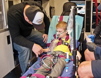
COVID-19 News: Italian researchers from the Pediatric Cardiology Unit at Padua University and the Department of Cardiac Thoracic Vascular Sciences and Public Health at Padua Hospital University-Italy are warning after documenting two clinical cases studies, that unique and usual forms of SARS-CoV-2-related arrhythmias known as Fascicular Tachycardia were materializing in children!

Fascicular tachycardia is a distinct subgroup of idiopathic ventricular tachycardias (VT) that may be confused with either typical (VT) or supraventricular tachycardia (SVT). Fascicular tachycardia has specific electrocardiographic features and therapeutic options. It is characterized by relatively narrow QRS complex and right bundle branch block pattern. The QRS axis depends on which fascicle is involved in the re-entry.
To date, research on heart rhythm disorders in children affected by COVID-19 infection is quite lacking.
Past
COVID-19 News coverages have already shown that SARS-CoV-2 infections can cause arrhythmia in Post COVID individuals.
https://www.thailandmedical.news/news/breaking-new-york-study-finds-that-sars-cov-2-virus-damages-heart-pacemaker-cells-and-causes-arrhythmias
https://www.thailandmedical.news/news/arrhythmia-university-of-pennsylvania-researchers-say-that-critical-covid-19-patients-are-10-times-more-likely-to-develop-cardiac-arrest
https://www.thailandmedical.news/news/warning-numerous-studies-are-showing-that-mild-symptomatic-sars-cov-2-infections-can-lead-to-a-variety-of-cardiac-issues,-some-with-fatal-outcomes
https://www.thailandmedical.news/news/latest-covid-19-news-sars-cov-2-causes-heart-muscle-cells-cardiomyocytes-to-fuse-and-disrupts-heart-s-electrical-rhythm
https://www.thailandmedical.news/news/covid-19-news-italian-study-shockingly-discovers-that-all-exposed-to-the-sars-cov-2-virus-have-an-additional-90-percent-risk-of-developing-heart-failu
The study team reported of two clinical case studies involving an infant and a congenital heart disease (CHD) teenager with a pacemaker presented with fascicular tachycardia and atrial flutter, respectively, during COVID-19 pauci-symptomatic infection.
&
lt;br />
Though the hemodynamic condition was always stable, the self-resolving trend of the atrial flutter and progressive resolution of the ventricular tachycardia occurred in conjunction with the negativization of the swab.
Such unique tachyarrhythmias have been reported as a form of potential arrhythmic complication during active pauci-symptomatic COVID-19 infection for the first time ever.
The study findings were published in the peer reviewed journal: COVID.
https://www.mdpi.com/2673-8112/3/2/14
These two clinical cases studies represent the potential threat that COVID-19-associated arrhythmia cases pose to children.
Case One:
A male infant aged about 10-months was admitted to the pediatric emergency room after 24 hours of agitation and inconsolable crying without the presence of fever, gastrointestinal, or respiratory problems.
Initial l examination revealed that the infant's hemodynamic status was stable, and his blood pressure was typical for his age, but his heart rhythm exhibited tachycardia.
The attending physicians reported no cardiac pericardial rubs or murmurs.
Neither gastrointestinal nor respiratory pathological signals were identified.
However, as per the echocardiogram (ECG), 220 beats per minute (bpm) of sustained ventricular tachycardia were detected. There was no evidence of congenital cardiac disease, coronary dilatation, or ventricular dysfunction on the ECG. The newborn also had no family history of rhythm abnormalities or sudden death.
The nasopharyngeal swab collected pre-admission was COVID-19-positive, although all other blood tests, including inflammatory testing and troponin, were negative.
Subsequent SARS-CoV-2 serology revealed a high immunoglobulin (Ig)-M antibody titer. Even though a few paroxysms of tachycardia were still detected in the subsequent four days, continuous intravenous amiodarone treatment was beneficial.
The intravenous antiarrhythmic treatment was matched with oral somministration until amiodarone was administered entirely orally.
The arrhythmia was effectively managed, with no ventricular tachyarrhythmia recurrence reported. After five days, a second echocardiogram revealed normal systolic and diastolic biventricular function as well as normal coronary artery size. After 21 days of recovery employing oral antiarrhythmic medication with amiodarone, the patient was released with a stable sinus rhythm.
Case Two:
A female adolescent aged 16 years with a congenital heart disease atrioventricular septal defect (AVSD) that was surgically corrected along with a dual chamber pacemaker used for post-surgical total AV block experienced palpitations two days prior to admission in the pediatric emergency room.
It was reported that at the commencement of symptoms, the device's remote control revealed atrial flutter onset two days prior. It was noted that this supraventricular tachyarrhythmia had never been recognized during prior periodic post-meridiem (PM) controls via telemedicine.
The female teenager arrived at the emergency department with a stable hemodynamic condition and no symptoms, while the ECG revealed an atrial flutter 2:1 at a 120 bpm heart rate.
Again, the nasopharyngeal swab obtained before patient admission was COVID-19-positive, although all other blood tests were negative.
It was found that C-reactive protein (CRP), and all other tests for inflammation, as well as troponin, tested negative. The ECG revealed no evidence of atrioventricular valve insufficiency, coronary dilation, or ventricular dysfunction.
Anticoagulant medication with subcutaneous heparin was administered without taking into account an antiarrhythmic treatment.
It was found that after 15 days in quarantine, the patient's nasopharyngeal swab tested COVID-19-negative. The instrument remote control revealed the spontaneous restoration of sinus rhythm in accordance with the swab's negativization. In the following medium- and short-term follow-ups, the study team identified no supraventricular arrhythmias.
Pediatric Arrhythmias
Both these cases represent the first time these tachyarrhythmias have been documented as probable arrhythmic consequences with active paucisymptomatic SARS-CoV-2 infection.
The self-resolving pattern of the AFL and the eventual fascicular tachycardia resolution was reported in concert with swab negativization.
Certain past studies and reviews have reported a correlation between electrophysiologic abnormalities and COVID-19 in children.
One past study described a pediatric population affected by COVID-19 infection with low prevalence of significant arrhythmias (NSVT or AT) in normal heart.
https://pubmed.ncbi.nlm.nih.gov/32621881/
Another a rare case report described that during the acute phase of symptomatic COVID-19 infection, a 9-day-old girl presented with aberrant supraventricular tachycardia, which was correctly diagnosed and then effectively treated by overdrive through a transesophageal pacing.
https://pubmed.ncbi.nlm.nih.gov/33931425/
To date, no studies or case reports have described an atrial flutter in congenital heart disease during a COVID-19 infection, especially in regard to pediatric population.
One documented case report only about an adult patient with a previously healthy heart who had been affected by severe SARS-CoV-2 infection that was complicated by cardiovascular involvement, especially an arrhythmic one.
https://www.jacc.org/doi/abs/10.1016/j.jaccas.2020.11.015
This adult patient presented an atrial flutter with high ventricular response rate that further compromised the cardiac pump function and the critical respiratory situation. The antiarrhythmic therapy failed, and catheter ablation had to be performed, despite the patient’s critically ill condition. The procedure was effective in terms of the stable restoration of sinus rhythm and respiratory conditions.
However, unlike the above adult case, the study team did not have to perform any medical therapy or invasive procedure during the acute phase because the girl was completely asymptomatic both for cardiac hemodynamic involvement and respiratory involvement, despite the cardiac heart disease.
The hypothesis of concomitant myocarditis was ruled out by analyzing the primary data collected from cardiac objectivity, the negative blood test, and the normal biventricular function in both of the patients. The myocardial involvement during the acute phase of SARS-CoV-2 infection is well known, but these two patients basically had arrhythmic complications of the acute COVID-19 infection, which were then followed by stable restoration of sinus rhythm.
https://pubmed.ncbi.nlm.nih.gov/33761041/
This is the first time that researchers have recorded this type of supraventricular tachycardia as it occurred spontaneously in a CHD pediatric patient with stable hemodynamic condition.
Such a type of tachyarrhythmia is typically related as a potential arrhythmic consequence during post-surgical follow-up of this CHD; nevertheless, the hemodynamic instability has not been previously reported in this case or from any other potential causes as well. The temporal correlation between the duration of this supraventricular tachycardia and the microbiological positivization might suggest the role of this virus as trigger factor of cardiac arrhythmia.
The varied and broad spectrum of ventricular tachyarrhythmias (VTs) has been already reported as arrhythmic involvement which is related to hemodynamic deterioration in critical COVID-19 disease, but no fascicular tachycardia has been documented either in adult or in pediatric populations.
Alarmingly, considering the quite rare manifestation of this VT during the first decade of life of a healthy child, it is undoubtedly even rarer to record a fascicular tachycardia in a 10- month-old baby with normal heart and stable hemodynamic condition.
This is the first time ever that this idiopathic ventricular tachycardia has been registered in a healthy infant who also had a documented SARS-CoV-2 infection.
This raises the question as to whether a SARS-CoV-2 infection could be a co-factor of fascicular tachycardia as in these cases. The study team believes that this might be a possibility, which is supported by the good control of the heart rhythm on antiarrhythmic therapy after the negativization of the swab.
For the latest
COVID-19 News, keep on logging to Thailand Medical News.
Read Also:
https://www.thailandmedical.news/news/warning-covid-19-clinical-care-experimental-drugs-being-tried-for-covid-19-can-cause-dangerous-abnormal-heart-rhythms
