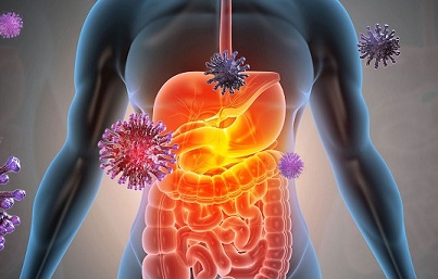
COVID-19 Research: American Study Confirms SARS-CoV-2 Infection Via The Human Gastrointestinal Tract And Also Explains The Causes Of Diarrhea.
Source: COVID-19 Research May 02, 2021 4 years, 7 months, 1 week, 5 days, 8 hours, 27 minutes ago
COVID-19 Research: A new study by researchers from Johns Hopkins University School of Medicine in Baltimore, Maryland, and the University of New Mexico Health Sciences Center in Albuquerque not only confirms the infection of SARS-CoV-2 via the gastrointestinal tract but also helps to explain the occurrence of diarrhea in many infected with the COVID-19 disease.

It has been found that diarrhea occurs in 2-50% of cases of COVID-19 (∼8% is average across series). The diarrhea does not appear to account for the disease mortality and its contribution to the morbidity has not been defined, even though it is a component of
Long COVID or post-infectious aspects of the disease.
Not much is known about the pathophysiologic mechanism of the diarrhea. In order to understand the pathophysiology of COVID-19 diarrhea, the study team exposed human enteroid monolayers obtained from five healthy subjects and made from duodenum, jejunum, and proximal colon to live SARS-CoV-2 and virus like particles (VLPs) made from exosomes expressing SARS-CoV-2 structural proteins (Spike, Nucleocapsid, Membrane and Envelope).
The live SARS-CoV-2 coronavirus was exposed apically for 90 min, then washed out and studied 2 and 5 days later.
The study found that SARS-Cov-2 was taken up by enteroids and live virus was present in lysates and in the apical basolateral media of polarized enteroids 48 hours after exposure.
This is the first demonstration of basolateral appearance of live virus after apical exposure. High vRNA concentration was detected in cell lysates and in the apical and basolateral media up to 5 days after exposure.
Also two days after viral exposure, cytokine measurements of media showed significantly increased levels of IL-6, IL-8 and MCP-1. Also two days after viral exposure, mRNA levels of ACE2, NHE3 and DRA were reduced but there was no change in mRNA of CFTR. NHE3 protein was also decreased.
Live viral studies were mimicked by some studies with VLP exposure for 48 h. VLPs with Spike-D614G bound to the enteroid apical surface and was taken up; this resulted in decreased mRNA levels of ACE2, NHE3, DRA and CFTR.
VLP effects were determined on active anion secretion measured with the Ussing chamber/voltage clamp technique. S-D614G acutely exposed to apical surface of human ileal enteroids did not alter the short-circuit current (Isc).
However, VLPS-D614G exposure to enteroids that were pretreated for ∼24 h with IL-6 plus IL-8 induced a concentration dependent increase in Isc indicating stimulated anion secretion, that was delayed in onset by ∼8 min. The anion secretion was inhibited by apical exposure to a specific calcium activated Cl channel (CaCC) inhibitor (AO1) but not by a specific CFTR inhibitor (BP027); was inhibited by basolateral exposure to the K channel inhibit clortimazole; and was prevented by pretreatment with the calcium buffer BAPTA-AM. 5) The calcium dependence of the VLP-induced increase in Isc was studied in Caco-2/BBe cells stably expressing the genetically encoded Ca2+ sensor GCaMP6s. 24 h pretreatment with IL-6/IL-8 did not alter intracellular Ca2+. However, in IL-6/IL-8 pretreated cells, VLP S-D614G caused appearance of Ca2+waves and an overall increase in i
ntracellular Ca2+ with a delay of ∼10 min after VLP addition.
The study team concluded that the diarrhea of COVID-19 appears to an example of a calcium dependent inflammatory diarrhea that involves both acutely stimulated Ca2+ dependent anion secretion (stimulated Isc) that involves CaCC and likely inhibition of neutral NaCl absorption (decreased NHE3 protein and mRNA and decreased DRA mRNA).
The study findings were published on a preprint server and are currently being peer reviewed.
https://www.biorxiv.org/content/10.1101/2021.04.27.441695v1
The study findings provided important insights into the pathophysiology of diarrhea that occurs in some cases of coronavirus disease 2019 (COVID-19).
The study team says the “COVID-19 diarrhea” that may develop following infection with the causative agent severe acute respiratory syndrome coronavirus 2 (SARS-CoV-2) is the first example of viral diarrhea that is dependent on the inflammatory response that occurs as part of the disease.
The study team has shown that one change in the intestinal transport process is the inhibition of two proteins (NHE3 and DRA) that enable neutral sodium chloride (NaCl) absorption ie the primary way that sodium is absorbed from the intestine in between meals.
It is known that the secretion of chloride is stimulated by activating the calcium-activated chloride channel (CaCC) by a mechanism dependent on increased intracellular calcium ions (Ca 2+).
Dr Olga Kovbasnjuk from the University of New Mexico Health Sciences Center told Thailand Medical News, “The diarrhea of COVID-19 appears to be an example of a calcium-dependent inflammatory diarrhea that involves both acutely stimulated Ca2+ dependent anion secretion that involves CaCC and likely inhibition of neutral NaCl absorption.”
To date, the number of recognized clinical manifestations of COVID-19 continues to increase, including the persistent manifestations seen in prolonged disease or “Long COVID.”
It has been observed that gastrointestinal (GI) manifestations such as diarrhea present in the early stages of the disease, but can occur throughout the course of the disease, including during the prolonged phase.
So far these GI manifestations are thought to occur as a result of direct luminal rather than systemic contact between SARS-CoV-2 and the GI tract.
Dr Kovbasnjuk added, “SARS-CoV-2 can be recovered from the lumen of the intestine-where it binds and replicates in human enterocytes.”
Key gaps in understanding of the effects SARS-CoV-2 has on the intestine include the intestinal sites affected, the mechanisms through which the virus causes diarrhea and whether the inflammatory response that occurs in COVID-19 contributes to producing diarrhea.
Dr Kovbasnjuk said, “Also unknown is the role of the GI tract in the many clinical aspects of SARS-CoV-2 infection, including viral replication and disease progression.”
The study team exposed a human enteroid model composed of enteroid monolayers from five healthy individuals to live SARS-CoV-2 as well as virus-like particles (VLPs) made from exosomes expressing the viral spike, nucleocapsid, membrane, and envelope proteins.
Interestingly forty-eight hours after initial exposure, live SARS-CoV-2 and viral replication were observed in enterocytes, supporting prolonged viral replication. The live virus was also present in apical and basolateral media.
Furthermore a high concentration of viral RNA was also detected in the apical and basolateral media up to five days following exposure.
This is the first evidence of systemic entry of SARS-CoV-2 via the GI tract.
As a result of the intestinal mucosal capillaries and lymphatics are found at the base of the epithelial cells, where live virus emerges from the basolateral surface of the enterocytes, these results provide the first evidence that intestinal SARS-CoV-2 infection may be involved in systemic entry of virus via the GI tract.
Dr Kovbasnjuk added, “Basolateral viral particles (live or not) might contribute to the widespread damage that is part of COVID-19.However, since systemic infection as part of COVID-19 is not felt to be an important part of the pathophysiology of this disease, the clinical relevance of this observation remains undefined.”
Also two days after exposure to the live virus, the enteroid media also showed significant increases in levels of the inflammatory cytokines interleukin (IL)-6, IL-8 and monocyte chemoattractant protein-1.
At the same time, levels of NHE3 and DRA, which enable NaCl absorption, were also reduced, with levels of NEH3 reduced at both the mRNA and the protein levels.
It was found that forty-eight hours following exposure of the enteroid model to VLPs, particles expressing the spike protein were taken up at the enteroid apical surface, which resulted in decreased mRNA levels of NHE3 and DRA. mRNA levels of the cystic fibrosis transmembrane conductance regulator (CFTR) and the human receptor for the viral spike protein – angiotensin-converting enzyme 2 (ACE2) – were also reduced.
Importantly the VLPs induced stimulated secretion of Cl via a Ca2+ dependent mechanism that was delayed with a time course that fitted the time for the VLP-induced increase in intracellular Ca2+. This VLP effect consistently required both binding and uptake into the enterocyte.
The study team found that the VLP-induced anion secretion could be inhibited by apical exposure to a specific calcium-activated Cl channel (CaCC) inhibitor (AO1), but not by exposure to a specific CFTR inhibitor (BP027). Anion secretin was also inhibited by basolateral exposure to the potassium channel inhibitor clotrimazole and was completely prevented by pre-treatment with the calcium buffer BAPTA-AM.
The study team concluded, “The mechanism of COVID-19 diarrhea appears to require the molecular interplay among inflammatory cytokines, the viral receptor, and several apical and basolateral ion transport proteins as well as activation of intracellular Ca2+ signaling. Even though the characterization of the changes in Na and Cl transport which occur throughout COVID-19 is incomplete, the results obtained already suggest potential drug targets while mechanistic insights may be relevant to SARS-CoV-2 effects on other epithelial cells.”
Please help to donate to sustain this website and other sites we manage and also all our research initiatives and community projects. https://www.thailandmedical.news/p/sponsorship
For the latest
COVID-19 Research, keep on logging to Thailand Medical News.
