COVID-19 Research: Study Finds That SARS-CoV-2 ORF3c Protein Localizes To Mitochondria, Inhibiting Innate Immunity By Restricting IFN-Β Production!
Source: COVID-19 Research - ORF3c - Mitochondria Nov 26, 2022 2 years, 4 months, 2 weeks, 4 days, 18 hours, 57 minutes ago
COVID-19 Research: A new international led by researchers from University of Cambridge - UK has found that the SARS-CoV-2 ORF3c protein localizes to mitochondria, inhibiting innate immunity by restricting IFN-Β production!

The study also involved scientists from University of Manchester - UK, University Medical Center Göttingen - Germany, University of Liverpool, University of Bristol, Broad Institute of MIT and Harvard - USA and University of Helsinki - Finland.
The SARS-CoV-2 genome encodes a multitude of accessory proteins. Utilizing comparative genomic approaches, an additional accessory protein, ORF3c, has been predicted to be encoded within the ORF3a sgmRNA.
The expression of ORF3c during infection has been confirmed independently by ribosome profiling. Despite ORF3c also being present in the 2002-2003 SARS-CoV, its function has remained unexplored.
The
COVID-19 Research team showed that ORF3c localizes to mitochondria during infection, where it inhibits innate immunity by restricting IFN-β production, but not NF-κB activation or JAK-STAT signaling downstream of type I IFN stimulation.
The study findings showed that ORF3c acts after stimulation with cytoplasmic RNA helicases RIG-I or MDA5 or adaptor protein MAVS, but not after TRIF, TBK1 or phospho-IRF3 stimulation. ORF3c co-immunoprecipitates with the antiviral proteins MAVS and PGAM5 and induces MAVS cleavage by caspase-3.
The study findings reveal an uncharacterized mechanism of innate immune evasion by this important human pathogen.
The study finings were published on a preprint server and are currently being peer reviewed.
https://www.biorxiv.org/content/10.1101/2022.11.15.516323v2
This is the first study involving the functional analysis of the severe acute respiratory syndrome coronavirus 2 (SARS-CoV-2) open reading frame 3c (ORF3c) protein.
The study team previously identified ORF3c using comparative genomics to be conserved among Sarbecoviruses.
https://pubmed.ncbi.nlm.nih.gov/32667280/
Subsequent, ribosome profiling studies have confirmed ORF3c expression during SARS-CoV-2 infection. ORF3c is also present in severe acute respiratory syndrome coronavirus 1 (SARS-CoV); however, the function of ORF3c needs to be better characterized and requires further investigation.
The study team hypothesized that the protein is expressed through ribosomal leaky scanning on ORF3a subgenomic messenger ribonucleic acid (sgmRNA), similar to ORF-7b and-9b of severe acute respiratory syndrome coronavirus 1 (SARS-CoV-1).
In order to test this hypothesis and to measure the protein expression levels, an expression cassette extending from the 5′ end of ORF3a sgmRNA transcript until the final coding nucleotide of the protein was created.
Next, GenBank sequence records of ORF3c sequences were inspected. Multiple mutants were created that a
blated the protein's initial and subsequent AUG codons, ORF3a AUG codons, or ORF3c AUG. Plasmid deoxyribonucleic acid (DNA) templates were transcribed in vitro by T7 RNA polymerase, and the transcripts were transfected in Vero cells.
To further investigate whether ORF3c was a transmembrane protein, transfection experiments were performed using Vero E6 cells, ORF3c-HA was overexpressed, and the cells were subjected to subcellular fractionation analysis.
Next, the nuclear, membranous, and cytoplasmic fractions were immunoblotted for ORF3c-HA detection. To evaluate ORF3c multimerization ability, ORF3c-HA and ORF3c-FLAG were co-transfected in the human embryonic kidney (HEK)293T cells, and immunoprecipitation analysis was performed.
In order to assess the membrane topology of ORF3c, the protein was modified to enable identification of ER import, and an OPG2 tag was incorporated at the C- or N-terminal ends. Radiolabelled ORF3c was synthesized, and the endoplasmic reticulum (ER) membranes of OPG2-tagged ORF3c variants were subjected to endoglycosidase H treatment to detect N-glycosylation.
ORF3C insertion ability into mitochondrial membranes was investigated based on transfection experiments using HEK293T cells and Vero cells, visualized by immunofluorescence microscopy after clear membrane staining.
Next, the immunoprecipitated ORF3c-HA proteins was analyzed by LC-MS (liquid chromatography and tandem mass spectrometry) to identify host interaction patterns. Further, the team investigated whether ORF3c could cause innate immune dysregulation.
The study team queried 12,906,225 sequences of SARS-CoV-2 from the GISAID (global initiative on sharing all influenza data) database to study the dynamics and appearance of ORF3c variants.
It was found that ORF3c, a conserved and small protein, overlapped the ORF3a protein in the +1 reading frame and coincided precisely with an area of significantly high synonymous site conservation in ORF3a-frame codons, indicating that the ORF3c gene was a functional SARS-CoV-2 gene.
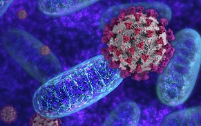
Importantly, ORF3c was found to localize in the outer mitochondrial membrane during SARS-CoV-2 infections, where the protein blocked innate immune pathways by limiting interferon-beta (IFN-β) expression, but not Janus kinase/signal transducers and activators of transcription (JAK-STAT) signaling or nuclear factor kappa B (NF-κB) activation.
The the morphology of mitochondria was however not altered by ORF3c presence in the SARS-CoV-2-infected cells. ORF3c acted after being stimulated by cytoplasmic ribonucleic acid helicase MDA5 (melanoma differentiation-associated gene 5) or RIG-I (retinoic acid-inducible gene I) or the MAVS (mitochondrial antiviral-signalling) adaptor protein, but not post-stimulation of TBK (TANK-binding kinase), TRIF (TIR-domain-containing adapter-inducing interferon-β), or phospho-interferon regulatory factor 3 (IRF3).
The ORF3c co-immunoprecipitated with the PGAM5 (phosphoglycerate mutase 5) and MAVS antiviral proteins and induced caspase 3-mediated cleavage of MAVS. By the leaky scanning hypothesis, mutation of the ORF3a AUGs doubled the expression of ORF3c.
Hence, nearly half of the pre-initiation complexes scanned past the first and second AUGs of ORF3a sgmRNA and initiated at the next AUG downstream for ORF3c translation.
It was also found that ORF3c did not multimerize and was not likely to form ionic channels.
It was observed that ORF3C imported and synthesized in vitro was shielded from added protease enzymes and was resistant to sodium carbonate buffer extraction, indicating that the ORF3c protein could stably integrate into ER membranes in vitro.
It was found that both terminals of OPG2-tagged ORF3c showed N-glycosylation and, therefore, could be translocated into ER microsome lumens. Interestingly, analyzing ORF3c using semi-permeabilized Henrietta Lacks (HeLa) cells, the degree of N-glycosylation was considerably reduced. In contrast, the total ORF3c protein signal was elevated, indicating that ORF3c might also be targeted to other cell organelles.
Significantly, ectopic ORF3c expression showed co-localization with endogenously expressed Tom70.
The mitochondrial membrane localization of the protein remained unaltered even after two days of transfection. Seven ORF3c variants were detected with ≥0.1% abundance, including the original WT variant and mutated L21F/R36I, R36I, Q5Y, S22L, K17E, and the delta PTC (premature termination codon) variant.
The study findings showed that the ORF3c protein is a tail-anchored transmembrane protein that inserts into the mitochondrial outer membrane, interacting with PGAM5 and MAVS to decrease IFN-β signaling.
The study findings could explain certain symptoms and conditions seen in COVID-19 and also Long COVID and could also help in the development of therapeutics to deal with these issues.
For the latest
COVID-19 Research, keep on logging to Thailand
Medical News.
