Diabetic Retinopathy Could Be Treated By Vitamin A Analog According To Researchers From The University Of Oklahoma
Source: Diabetic Retinopathy Jun 12, 2020 5 years, 7 months, 1 week, 8 hours, 53 minutes ago
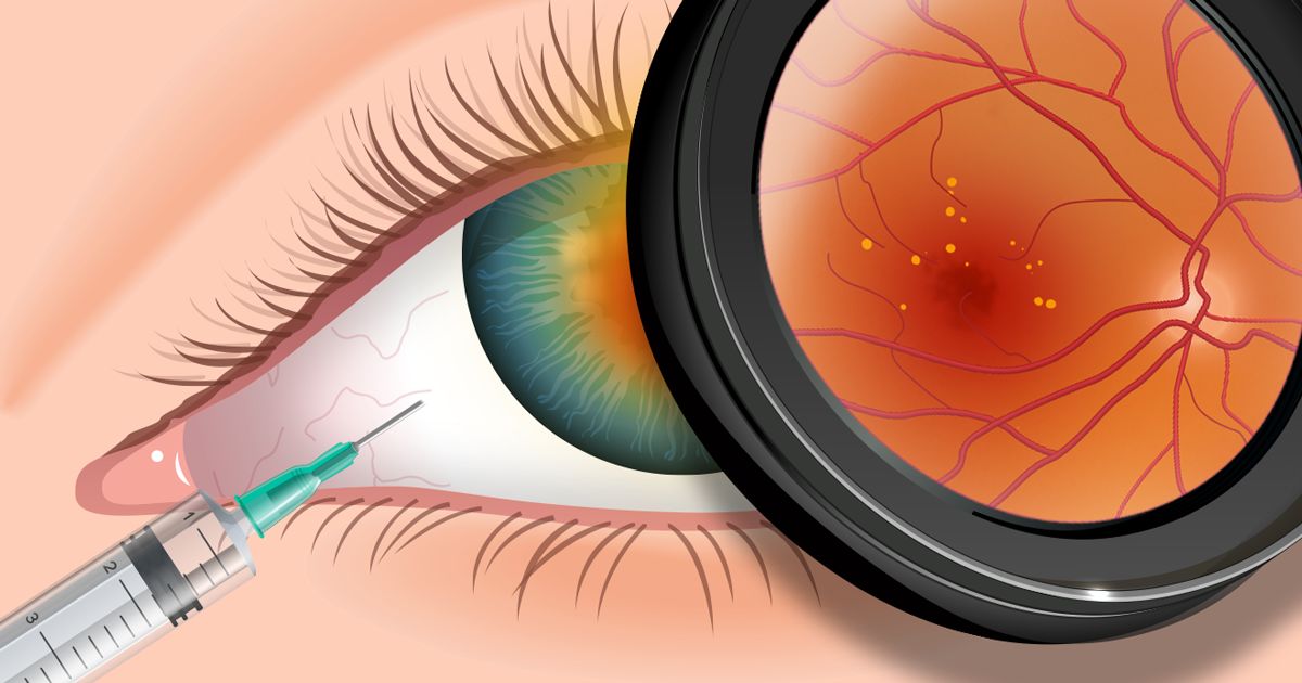
Diabetic retinopathy is a typical complication of diabetes and a leading cause of blindness in the world. A new research by scientist from University Of Oklahoma show that visual function in diabetic mice was significantly improved after treatment with a single dose of visual chromophore 9-cis-retinal, a vitamin A analog that can form a visual pigment in the retina cells, thereby producing a light sensitive element of the retina.

The research study was published in the
American Journal of Pathology.
https://ajp.amjpathol.org/article/S0002-9440(20)30146-2/pdf
Dr Gennadiy Moiseyev, from the department of Physiology, University of Oklahoma Health Sciences Center and lead investigator told Thailand Medical News, "In an earlier study we found that diabetes causes vitamin A deficiency in the retina, which results in deterioration of vision, even before any vascular changes can be seen. That finding led to the assumption that early changes in vision in diabetes are probably caused by vitamin A deficiency in the retina."
The researchers hypothesized that treating diabetic mice with 11-cis-retinal could rescue visual function. They investigated its effect in Akita mice, a genetic model of type 1 diabetes, by measuring electroretinogram (ERG) responses, retinal oxidative stress, and neuronal apoptosis (cell death). ERG was performed on two groups of three-month-old Akita mice and one group of non-diabetic control mice matched for age and genetic background. One group of Akita mice was treated with 9-cis-retinal and the other with a vehicle solution. Average blood glucose concentrations and body weights of the mice were measured monthly during the study. Akita mice showed high glucose concentrations throughout the study. ERG recordings and rhodopsin assay were performed two hours after 9-cis-retinal injection, whereas assessment of cell death by ELISA and TUNEL assay were both performed 24 hours after the injection.
The study results showed that the visual function in diabetic mice improved significantly after treatment with the single dose of 9-cis-retinal. In addition, researchers reported that the treatment reduced oxidative stress in the retina, decreased retina cell death and retina degeneration, and improved visual function.
Dr Moiseyev added, "This research supports our novel hypothesis that diabetes-induced disturbance of the vitamin A metabolism in the eye is responsible for reduced visual function in early stages of diabetic retinopathy,"
He further added, "Currently, there is no available therapy to prevent the development of the retinal complication in patients suffering from diabetes. This study suggests that the delivery of visual chromophore to the diabetic eye may represent a potential therapeutic strategy for the early stages of diabetic retinopathy to prevent vision loss in patients with diabetes."
Conventionally, diabetic retinopathy was considered a disease caused by the pathology of blood vessels in the retina, whereby light-sensitive tissue at the back of the eye (fundus) becomes damaged. Patients with diabetes often experience functional deficits in dark adaptation, contra
st sensitivity, and color perception before any microvascular pathologies are detected on the eye fundus.
Newer data however underscore the importance of vitamin A for normal visual function. It serves as a precursor for light-sensitive 11-cis-retinal, the chromophore of visual pigments that can produce a light-sensitive protein in the retina.
The team is planning to start human clinical trials as early the later part of the year, sometime in September.
For more on
diabetic retinopathy, keep on logging to Thailand Medical News.
