For All The Latest Medical News, Health News, Research News, COVID-19 News, Dengue News, Glaucoma News, Diabetes News, Herb News, Phytochemical News, Cardiology News, Epigenetic News, Cancer News, Doctor News, Hospital News
First Implantable Magnet Resonance Detector Brain Probe Developed By Neuroscientists To Measure Neuronal Activity
Source: Thailand Medical News Dec 01, 2019 4 years, 11 months, 2 weeks, 6 days, 14 hours, 5 minutes ago
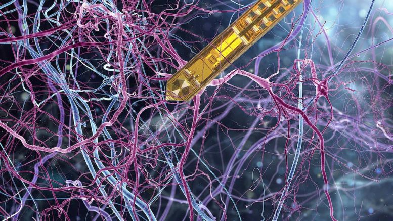
German and Swiss neuroscientists and electrical engineers have developed a highly sensitive implant that enables to probe brain physiology with unparalleled spatial and temporal resolution. They introduce an ultra-fine needle with an integrated chip that is capable of detecting and transmitting nuclear magnetic resonance (NMR) data from nanoliter volumes of brain oxygen metabolism. The breakthrough design will allow entirely new applications in the life sciences.
Illustration of the tiny MRT needle in the brain tissue. Credit: whitehoune MPI f. Biological
Cybernetics, University of Stuttgart. Compositing: Martin Vötsch (design-galaxie.de).
The team of researchers led by Dr Klaus Scheffler from the Max Planck Institute for Biological Cybernetics and the University of Tübingen as well as by Jens Anders from the University of Stuttgart identified a technical bypass that bridges the electrophysical limits of contemporary brain scan methods. Their development of a capillary monolithic nuclear magnetic resonance (NMR) needle combines the versatility of brain imaging with the accuracy of a very localized and fast technique to analyze the specific neuronal activity of the brain.
Principal investigator Dr Klaus Scheffler told Thailand Medical News, “The integrated design of a nuclear magnetic resonance detector on a single chip supremely reduces the typical electromagnetic interference of magnetic resonance signals. This enables neuroscientists to gather precise data from minuscule areas of the brain and to combine them with information from spatial and temporal data of the brain´s physiology. With this method, we can now better understand specific activity and functionalities in the brain.”
Dr Scheffler says that their invention may unveil the possibility of discovering novel effects or typical fingerprints of neuronal activation, up to specific neuronal events in brain tissue. “Our design setup will allow scalable solutions, meaning the possibility of expanding the collection of data from more than from a single area but on the same device. The scalability of our approach will allow us to extend our platform by additional sensing modalities such as electrophysiological and optogenetic measurements,“ adds the second principal investigator Jens Anders.
The researchers are very confident that their technical approach may help demerge the complex physiologic processes within the neural networks of the brain and that it may uncover additional benefits that can provide even deeper insights into the functionality of the brain. With their primary goal to develop new techniques that are able to specifically probe the structural and biochemical composition of living brain tissue, their latest innovation paves the way for future highly specific and quantitative mapping techniques of neuronal activity and bioenergetic processes in the brain cells.
Reference: “A CMOS NMR needle for probing brain physiology with high spatial and temporal resolution” by Jonas Handwerker, Marlon Pérez-Rodas, Michael Beyerlein, Franck Vincent, Armin Beck, Nico
las Freytag, Xin Yu, Rolf Pohmann, Jens Anders and Klaus Scheffler, 25 November 2019, Nature Methods. DOI: 10.1038/s41592-019-0640-3