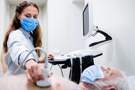
Lung ultrasound proves effective in identifying long-term post-COVID-19 lung damage
Nikhil Prasad Fact checked by:Thailand Medical News Team Sep 22, 2024 7 months, 2 weeks, 3 days, 7 hours, 22 minutes ago
Medical News: As we continue to navigate the aftermath of the COVID-19 pandemic, researchers worldwide are exploring ways to monitor and address long-term health impacts in those who survived the virus. One critical area of interest is the detection and management of lung damage that can persist long after the initial infection. Recent research has highlighted the potential of lung ultrasound (LUS) in detecting such damage, offering a non-invasive and cost-effective alternative to more conventional imaging techniques.
 Lung ultrasound proves effective in identifying long-term post-COVID-19 lung damage
In this
Medical News report, we’ll explore how a systematic review and meta-analysis conducted by a team of researchers from various medical institutions in Italy shed light on the use of lung ultrasound to detect lung sequelae in post-COVID-19 patients. The research evaluated the effectiveness of LUS compared to traditional methods like chest X-rays (CXR) and computed tomography (CT) scans in identifying lung fibrosis and other long-term changes. The findings could shape how clinicians manage post-COVID care for years to come.
The Study: A Closer Look at Lung Ultrasound and Post-COVID Damage
The study, conducted by researchers from the University of Bologna, Italy; "G. d’Annunzio" University of Chieti, Italy; and IRCCS Azienda Ospedaliero-Universitaria di Bologna-Italy, investigated the effectiveness of lung ultrasound in detecting fibrotic-like changes in post-COVID-19 patients. In their systematic review, the researchers analyzed data from multiple studies to determine how accurately LUS could identify lung parenchymal damage (the area of the lungs responsible for gas exchange), particularly when compared to CT scans, the current gold standard for lung imaging.
The study examines how lung ultrasound is emerging as a valuable tool for detecting interstitial lung disease (ILD) in post-COVID patients. It also reviews the study’s findings on sensitivity, specificity, and overall diagnostic accuracy, providing insights into the future of non-invasive diagnostics in post-COVID-19 care.
Key Findings: Sensitivity, Specificity, and Diagnostic Accuracy
The study analyzed data from three key research articles, each comparing lung ultrasound to CT scans in post-COVID-19 patients. Two models were used to assess the diagnostic performance of LUS: a low-threshold model and a high-threshold model for detecting B-lines (a sign of lung damage seen on ultrasound). The B-lines are a visual indication of lung parenchymal changes, with more B-lines indicating more severe damage.
Low-Threshold Model
In the low-threshold model, the sensitivity of LUS was found to be exceptionally high, reaching 98% (95% CI: 95 - 99%). This means that lung ultrasound was able to correctly identify 98% of the patients with fibrotic-like changes in their lungs. However, the specificity in this model was lower, at 54% (95% CI: 49 - 59%). Specificity refers to the ability of a test to correctly ide
ntify patients who do not have the condition. The lower specificity means that LUS, in this model, may falsely identify lung damage in patients who are actually healthy.
The Diagnostic Odds Ratio (DOR) for this model was 44.9, and the Area Under the Curve (AUC) of the Summary Receiver-Operating Characteristic (SROC) curve was 0.90. The DOR indicates the effectiveness of a diagnostic test, while the AUC represents the test’s overall accuracy. A value of 0.90 is considered excellent.
High-Threshold Model
In contrast, the high-threshold model displayed a sensitivity of 90% (95% CI: 85 - 94%) and a much higher specificity of 88% (95% CI: 84 - 91%). This suggests that when a higher threshold for detecting B-lines is used, the accuracy of lung ultrasound in correctly identifying patients without lung damage improves significantly. The DOR for this model was 50.4, with an AUC of 0.93, indicating even better diagnostic performance than the low-threshold model.
In both models, LUS demonstrated high sensitivity, meaning it could effectively detect fibrotic-like changes in the lungs of post-COVID-19 patients. However, the variation in specificity between the two models shows that higher thresholds for B-line detection offer better diagnostic accuracy.
Why Lung Ultrasound?
During the COVID-19 pandemic, lung ultrasound became a valuable tool in diagnosing acute pneumonia caused by SARS-CoV-2. The pandemic reinforced the importance of LUS in emergency settings, where quick, accurate diagnosis is essential. Lung ultrasound offers several advantages over traditional imaging methods like chest X-rays or CT scans. It is non-invasive, non-ionizing, portable, and less costly, making it a more accessible option for patients in various settings.
LUS has already been proven effective in diagnosing lung conditions like interstitial lung disease caused by other factors, including connective tissue diseases. Given its ability to detect changes in lung parenchyma with similar sensitivity to CT scans, LUS offers a promising approach to monitoring post-COVID lung health.
Additional Findings: Pleural Line Irregularities
The study also highlighted other significant ultrasound findings in post-COVID patients, particularly changes in the pleural line. The pleural line is the outer layer of the lungs, and abnormalities in this area can indicate lung damage. In one of the studies reviewed, 77.3% of patients with persistent lung damage had a thickened pleural line, compared to only 35.9% of patients without residual damage.
Fragmented pleural lines, where the pleural layer appears uneven or broken, were also noted in 7.8% of patients with lung sequelae, but none in those who had fully recovered. These findings underscore the utility of LUS in providing a comprehensive picture of lung health, going beyond B-lines to include other critical indicators of lung damage.
Conclusion: What This Means for Post-COVID-19 Care
Lung ultrasound is proving to be an effective, reliable tool for diagnosing long-term lung damage in post-COVID-19 patients. With its high sensitivity, especially in detecting fibrotic-like changes, LUS can be an essential component of post-COVID care. Its non-invasive nature and lower costs compared to CT scans make it an attractive option for regular follow-ups, especially for patients who have experienced significant lung involvement during their COVID-19 infection.
However, while lung ultrasound shows great promise, there are still some limitations. The study pointed out that LUS is highly dependent on the skill of the operator, meaning that variability in training and experience could affect the accuracy of results. Additionally, there is no standardized method for reporting LUS findings across studies, which makes it challenging to compare results and draw broad conclusions.
Nevertheless, the findings suggest that LUS could be used more widely in post-COVID monitoring, offering a more affordable and safer alternative to repeated CT scans. Future research should aim to standardize LUS protocols and further investigate its long-term utility in managing post-COVID lung conditions.
The study findings were published in the peer-reviewed Journal of Clinical Medicine.
https://www.mdpi.com/2077-0383/13/18/5607
For the latest COVID-19 News, keep on logging to Thailand
Medical News.
Read Also:
https://www.thailandmedical.news/news/diaphragm-muscle-atrophy-linked-to-long-term-fatigue-in-covid-19-survivors
https://www.thailandmedical.news/news/long-covid-patients-face-hidden-lung-problems-leading-to-persistent-breathlessness
