Source: Thailand Medical News Nov 20, 2019 6 years, 1 month, 2 weeks, 2 days, 8 hours, 37 minutes ago
An innovative biosensor technology developed by a Rutgers-led team has created better that may help lead to safe
stem cell therapies for treating Alzheimer’s and Parkinson’s diseases and other neurological disorders.
The new technology, which features a unique graphene and gold-based platform and high-tech imaging, monitors the fate of
stem cells by detecting genetic material (RNA) involved in turning such cells into brain cells (neurons), according to the study that was published in the journal Nano Letters.
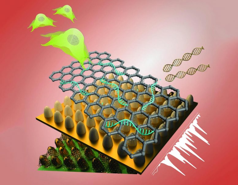
 This unique biosensing platform consists of an array of ultrathin graphene layers and gold nanostructures.
The platform, combined with high-tech imaging (Raman spectroscopy), detects genetic material (RNA) and
characterizes different kinds of stem cells with greater reliability, selectivity and sensitivity than today’s
biosensors. Credit: Letao Yang, KiBum Lee, Jin-Ho Lee and Sy-Tsong (Dean) Chueng
Typically
stem cells can become many different types of cells. As a result, stem cell therapy shows promise for regenerative treatment of neurological disorders such as Alzheimer’s, Parkinson’s, stroke and spinal cord injury, with diseased cells needing replacement or repair. But characterizing
stem cells and controlling their fate must be resolved before they could be used in treatments. The formation of tumors and uncontrolled transformation of
stem cells remain key barriers.
Dr KiBum Lee, a professor in the Department of Chemistry and Chemical Biology in the School of Arts and Sciences at Rutgers University-New Brunswick and senior author told
Thailand Medical News, “A critical challenge is ensuring high sensitivity and accuracy in detecting biomarkers indicators such as modified genes or proteins within the complex
stem cell microenvironment.Our technology, which took four years to develop, has demonstrated great potential for analyzing a variety of interactions in
stem cells.”
The research team’s unique biosensing platform consists of an array of ultrathin graphene layers and gold nanostructures. The platform, combined with high-tech imaging (Raman spectroscopy), detects genes and characterizes different kinds of
stem cells with greater reliability, selectivity, and sensitivity than today’s biosensors.
The researchers believes the technology can benefit a range of applications. By developing simple, rapid and accurate sensing platforms, Lee’s group aims to facilitate treatment of neurological disorders through
stem cell therapy.
Stem cells may become a renewable source of replacement cells and tissues to treat diseases including macular degeneration, spinal cord injury, stroke, burns, heart disease, diabetes, osteoarthritis, and rheumatoid arthritis, according to the National Institutes of Health.
The study’s co-lead
authors are Letao Yang and Jin-Ho Lee, postdoctoral researchers in Lee’s group. Rutgers co-authors include doctoral students Christopher Rathnam and Yannan Hou. A scientist at Sogang University in South Korea contributed to the study.
Reference: “Dual-Enhanced Raman Scattering-Based Characterization of Stem Cell Differentiation Using Graphene-Plasmonic Hybrid Nanoarray” by Letao Yang, Jin-Ho Lee, Christopher Rathnam, Yannan Hou, Jeong-Woo Choi and Ki-Bum Lee, 30 October 2019, Nano Letters. DOI: 10.1021/acs.nanolett.9b03402 