Nikhil Prasad Fact checked by:Thailand Medical News Team Nov 16, 2024 4 months, 3 weeks, 6 days, 3 hours, 29 minutes ago
Medical News: Researchers from the Medical University of Bialystok, Poland, have unveiled key insights into post-COVID-19 brain alterations using advanced imaging techniques. The study employed Vessel Wall Imaging (VWI) and other MRI methods to assess lingering neurological effects in patients recovering from COVID-19.
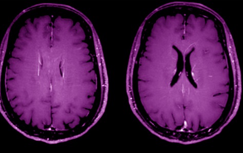
 Axial MIP reconstruction of the post-contrast 3D T1-weighted images at the level of
lateral ventricles shows bilateral perivascular enhancement along engorged deep medullary veins
Unraveling Post-COVID Brain Anomalies
COVID-19 is no longer just a respiratory ailment; its aftershocks ripple through various organs, especially the brain. This
Medical News report reveals how the study examined 43 participants - 23 post-COVID patients and 20 healthy controls - using cutting-edge MRI. The post-COVID group, six months into recovery, displayed a spectrum of changes linked to brain health and vessel integrity.
The researchers focused on Vessel Wall Imaging (VWI), an innovative MRI tool that captures intricate details of blood vessel walls, offering insights into underlying neurological complications. Results painted a vivid picture of the potential neurological toll of the pandemic.
Significant Findings in Post-COVID Brains
The imaging highlighted numerous abnormalities in post-COVID patients:
-Hyperintense Foci in Brain Regions: Post-COVID patients exhibited hyperintense lesions in white matter, the lower temporal lobes, and structures in the posterior cranial fossa, suggesting potential inflammatory or ischemic damage. These lesions were present in 87% of the patients.
-Engorgement and Perivascular Enhancements: Imaging revealed the engorgement of deep medullary veins and perivascular enhancement, indicating vessel inflammation in 13% of cases.
-Vessel Wall Abnormalities: Concentric thickening of vessel walls, often linked to inflammation, was observed in 30% of post-COVID patients, while atherosclerotic changes were found in nearly half.
-Cortical Atrophy and Ventricular Width: Marked cortical atrophy (83%) and a widened third ventricle - both linked to aging and brain health - were more prevalent in the post-COVID group.
Sinus and Hippocampal Changes: Mucosal thickening in the paranasal sinuses (65%) and alterations in hippocampus size pointed to broader systemic involvement.
Understanding the Broader Implications
These findings suggest that COVID-19 survivors, even months after infection, may face persistent neurological and vascular challenges. The vessel wall abnormalities, though milder than typical CNS vasculitis, highlight the virus's potential to trigger systemic inflammation. Early detection through VWI could pave the way for targeted interventions.
The
study also shed light on age-related trends. Older patients displayed more pronounced vessel wall changes and white matter abnormalities, possibly exacerbated by COVID-19.
Why Vessel Wall Imaging Stands Out
VWI stands apart by focusing on vessel walls rather than just luminal structures, making it invaluable for detecting early inflammatory changes. Researchers used a high-resolution 3T MRI scanner, ensuring superior image quality. This technique could revolutionize how we monitor post-viral syndromes like long COVID, guiding therapy to reduce future complications.
Conclusion
The research underscores that post-COVID brains are far from returning to baseline, even months after recovery. Persistent lesions and vascular changes necessitate vigilant monitoring, particularly in older populations. Advanced imaging like VWI might not only refine diagnoses but also improve outcomes by identifying high-risk patients early.
The study findings were published in the peer-reviewed Journal of Clinical Medicine.
https://www.mdpi.com/2077-0383/13/22/6884
For the latest COVID-19 News, keep on logging to Thailand
Medical News.
Read Also:
https://www.thailandmedical.news/news/zinc-sulfate-may-block-covid-19-brain-invasion
https://www.thailandmedical.news/news/how-salidroside-helps-protect-the-brain-after-stroke
