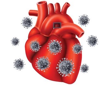
Research Finds That SARS-CoV-2 S And ORF-9B Proteins Alters Metabolic Profiles And Impairs Contractile Function In Cardiomyocytes!
SARS-CoV-2 Research - Spike And ORF-9B Proteins Impairs Contractile Function In Cardiomyocytes! Mar 14, 2023 2 years, 1 month, 6 days, 4 hours, 52 minutes ago
SARS-CoV-2 Research: Scientists from Clemson University, South Carolina-USA, Stanford University School of Medicine, California-USA, Southern Illinois University School of Medicine-USA, Shandong University, Jinan-China and University of Pittsburgh-USA have in a new study found that ectopic expression of SARS-CoV-2 S and ORF-9B proteins alters metabolic profiles and impairs contractile function in cardiomyocytes.

While, the impacts of COVID-19 on the cardiovascular system are known, the specific mechanisms involved are still unclear.
The study team conducted "pseudoviral infection" of SARS-CoV-2 subunits on human induced pluripotent stem cell-derived cardiomyocytes (CMs) to evaluate their toxic effects.
The study findings indicate that the expression of S and ORF-9B subunits significantly impaired the contractile function and altered metabolic profiles in human CMs.
Specifically, mitochondrial oxidative phosphorylation, membrane potential, and ATP production was observed two days after S and ORF-9B overexpression, as well as elevated levels of reactive oxygen species (ROS) induced by S subunits.
Two weeks after overexpression, the study team found increased glycolysis in the ORF-9B group.
Transcriptomic analysis showed that both S and ORF-9B subunits dysregulated signaling pathways associated with metabolism and cardiomyopathy, including upregulated genes involved in HIF-signaling and downregulated genes involved in cholesterol biosynthetic processes. Furthermore, the ORF-9B subunit enhanced glycolysis in the CMs.
Overall, the study findings shed light on the molecular mechanisms underlying SARS-CoV-2 subunit-induced metabolic alterations and cardiac dysfunctions in COVID-19 patients.
The study findings were published in the peer reviewed journal: Frontiers In Cell And Developmental Biology.
https://www.frontiersin.org/articles/10.3389/fcell.2023.1110271/full
COVID-19, caused by severe acute respiratory syndrome coronavirus 2 (SARS-CoV-2), is a respiratory disease that can be fatal. To date, it has been estimated that there have been about 2.5 billion cases of infection and about 37 million COVID-19 deaths globally not including excess deaths! Impact of COVID-19 on global public health and socio-economic development has been devastating.
The virus infects the host by binding the surface spike protein (S protein) to angiotensin-converting enzyme 2 (ACE2), which is the primary receptor for the virus. ACE2 is present in high levels in the lungs and heart, causing cardiovascular complications through the interaction with the S protein of the virus. However, the specific mechanisms behind the cardiovascular complications caused by individual SARS-CoV-2 subunits are not yet fully understood.
In addition, patients with pre-existing cardiovascular disease or risk factors may be more likely to experience severe symptoms.
While the respiratory system is the primary target of SARS-CoV-2, the virus can also cause damage to other organs. For instance, elevated levels of cardiac troponin I have been observed in hospitalized patients with COVID-19, indicating cardiac damage followin
g infection. Moreover, patients with pre-existing cardiovascular disease have a higher risk of mortality. This highlights the potential for SARS-CoV-2 infection to exacerbate underlying cardiac conditions and cause new cardiovascular complications, including myocardial injury, in some hospitalized patients.
Human stem cell-derived cardiomyocytes (CMs) are a valuable tool for investigating mechanistic and toxicological aspects of the cardiovascular system. To advance this research, we previously developed an approach that combines human stem cells with transcriptomics and epigenomics to identify transcriptional regulatory mechanisms involved in early CM differentiation and drug responses.
Recently, several studies have explored the impact of SARS-CoV-2 infection on iPSC-derived CMs. These investigations have revealed significant transcriptomic and functional remodeling, thereby improving our understanding of COVID-19 related cardiac risks using human-originated cellular models.
While it is known that SARS-CoV-2 nucleocapsid proteins are overexpressed in host cells following transfection, the specific role of each protein in inducing cardiac toxicity and functional failure remains unclear.
To address this gap, the study team conducted this study using "pseudoviral infection" of SARS-CoV-2 subunits in hiPSC-derived CMs to evaluate cardiac functions and metabolic profiles. Additionally, they employed genome-wide transcriptomics to investigate the underlying mechanisms responsible for the adverse impacts of SARS-CoV-2 subunits on CMs.
The impacts of coronavirus SARS-CoV-2 subunits on human iPSC-derived CMs were assessed in this research. Long-term infection with S and ORF-9B subunits resulted in metabolic changes and cardiac dysfunctions. A toxicogenomic analysis revealed that the dysregulation of metabolic and cardiomyopathy pathways occurred due to both S and ORF-9B subunits. Additionally, ORF-9B subunit augmented glycolysis, resulting in metabolic remodeling in the infected CMs.
The proper management of cardiometabolic health is crucial for maintaining optimal cardiac function, which entails meeting high ATP demands. While mature cardiomyocytes primarily rely on mitochondrial oxidative phosphorylation (OXPHOS) to produce ATP, cardiomyopathy patients may exhibit metabolic reprogramming, whereby the balance between glycolysis and mitochondrial OXPHOS shifts.
Moreover, chronic metabolic diseases such as diabetes and cardiovascular disease are closely linked to the immune system and metabolism. As a result, disturbances in metabolic homeostasis may result in systemic inflammatory responses, which may account for the increased mortality rates of patients with diabetes and cardiovascular disease who contract SARS-CoV-2. Such patients may also present severe inflammatory syndromes.
Furthermore, studies have demonstrated that SARS-CoV-2 infection can lead to the reprogramming of various nutrients' metabolism, including glucose, fatty acids, cholesterol, and glutamine.
A past
SARS-CoV-2 Research revealed that SARS-CoV-2 Nsp6 expression in the heart of Drosophila resulted in interaction with the host MGA/MAX complex (MGA, PCGF6, and TFDP1), which ultimately caused a rapid shift towards glycolysis. Moreover, the virus can influence the host cells' mitochondrial functions by releasing ORF proteins (e.g., ORF-9b) that localize in the host mitochondria.
In the current study, it was observed that similar metabolic reprogramming results from SARS-CoV-2 subunits in the human stem-cell-based system, with both ORF-9B and S proteins reducing mitochondrial OXPHOS levels and increasing glycolysis in CMs.
The study findings also showed that S protein augmented the mitochondrial ROS levels, and both ORF-9B and S proteins upregulated the HIF-1 signaling pathway and genes related to "cellular response to hypoxia," indicating a potential pathway where SARS-CoV-2 subunits induce hypoxia to activate HIF-1 signaling and elevate glycolysis.
This mechanism has been observed in monocytes after SARS-CoV-2 infection and is accompanied by increased ROS levels in mitochondria, leading to cytokine storms.
The study findings suggest a similar mechanism in CMs from SARS-CoV-2 infection. Additionally, viral infection affects cholesterol homeostasis, with decreased HDL cholesterol levels and higher triglycerides being demonstrated. SARS-CoV-2 infection is likely to activate sterol-regulatory element-binding protein 2 (SREBP-2), disrupting cholesterol biosynthesis or liver damages and lowered lipid metabolism.
The study findings showed that both ORF-9B and S downregulated genes involved in "cholesterol biosynthetic process," indicating changes in lipid metabolism in the heart following infection.
It was also found that some genes were significantly dysregulated in COVID-19 patients and showed similarity to previous clinical reports. For example, fibrinogen and CTGF, which are indicators for coagulation, fibrinolysis, and lung injury, were found to be elevated in COVID-19 patients.
Similarly, it was found that these genes were also upregulated in cardiomyocytes (CMs) after infection, indicating a similar pathological progression in the CMs.
Additionally, it was observed that the SARS-CoV-2 subunits can cause aberrant expression of cardiac genes.
The analysis of the enriched GO terms of the upregulated genes suggests that ORF-9B and S subunits can alter the transcriptional regulation in cardiac gene programs. Further investigation using other technologies, such as ATAC-seq, is necessary to understand additional mechanisms underlying transcriptional dysregulation.
It should be noted that this study utilized an in vitro system to gain insight into infectious diseases in vivo. This stem-cell-based system provides significant advances in the understanding of the cardiovascular system of COVID-19 patients. However, there are limitations to this system, such as the immaturity of the differentiated cardiomyocytes compared to the mature heart in vivo and a lack of spatial and cellular heterogeneity in the monolayer model compared to that of the heart. Therefore, future studies can benefit from the use of mouse models or 3D-cardiac organoids as an important complementary solution.
For the latest
SARS-CoV-2 Research, keep on logging to Thailand Medical News.
