Researchers Warn That Even Mild COVID-19 Can Cause Retinal Microvascular Changes of the Eyes
Nikhil Prasad Fact checked by:Thailand Medical News Team May 22, 2024 1 year, 4 months, 3 weeks, 3 days, 16 hours, 22 minutes ago
COVID-19 News: The COVID-19 pandemic has brought about significant global health challenges, affecting various organs beyond the respiratory system. Researchers from the Chinese Academy of Medical Sciences & Peking Union Medical College in Beijing, China, have recently conducted a comprehensive study to investigate the impact of mild COVID-19 infections on retinal microvasculature. This
COVID-19 News report delves into their findings, shedding light on the potential long-term implications of COVID-19 on eye health.
 Mild COVID-19 Can Cause Retinal Microvascular Changes of the Eyes
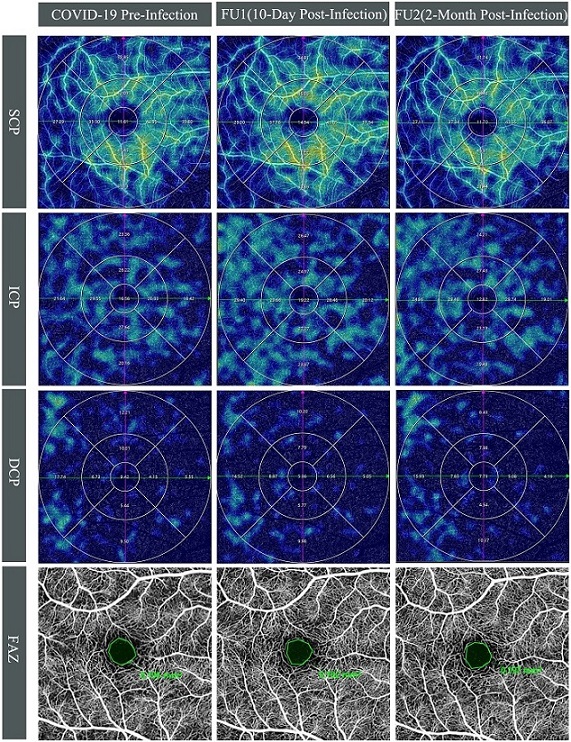
Longitudinal swept-source optical coherence tomography angiography (SS-OCTA) vessel density (VD) changes in the right eye of a 28-year-old male. Top three rows display SS-OCTA VD heatmaps of the superficial (SCP), intermediate (ICP), and deep capillary plexuses (DCP) across three time points: COVID-19 pre-infection, first follow-up (10-day post-infection), and second follow-up (2-month post-infection). Compared to pre-infection, an increase in VD was observed at first follow-up for SCP (from 33.49% to 35.75%) and ICP (from 22.23% to 25.28%), whereas VD approached pre-infection levels with SCP at 33.46% and ICP at 22.89% at the second follow-up. The DCP exhibited a marginal decline at the first follow-up (from 10.16% to 9.21%) and further decreased at the second follow-up (8.92%). Bottom row shows the foveal avascular zone (FAZ) at COVID-19 pre-infection, 10-day post-infection, and 2-month post-infection. The FAZ measurements remained relatively unchanged pre- and post-COVID-19 infection, with values of 0.195 mm² at baseline, 0.182 mm² at the first follow-up, and 0.197 mm² at the second follow-up.
Purpose of the Study
The primary objective of the study was to examine the longitudinal alterations in retinal microvasculature among patients who had contracted mild COVID-19. The researchers aimed to determine whether these changes were temporary or had lasting effects.
Methods
The study was conducted on a cohort of participants who had no prior history of COVID-19 infection. These individuals were recruited between December 2022 and May 2023 at the Peking Union Medical College Hospital in Beijing. Participants underwent thorough ophthalmologic examinations and fundus imaging, including color fundus photography, autofluorescence photography, swept-source optical coherence tomography (SS-OCT), and SS-OCT angiography (SS-OCTA).
If participants contracted COVID-19 during the study period, follow-up imaging was performed within one week and two months after their recovery to assess any changes in retinal microvasculature.
Participant Demographics
A total of 31 participants (61 eyes), with a mean age of 31.0 ± 7.2 years, were eligible for the study. All participants experienced mild COVID-19 infections within one
month of baseline data collection. The average period for the first follow-up was 10.9 ± 2.0 days post-infection, and the second follow-up occurred at 61.0 ± 3.5 days post-infection.
Retinal Microvasculature Findings
No clinical features of retinal microvasculopathy were observed during the follow-ups. However, SS-OCTA analysis revealed significant changes in retinal microvasculature. There was a notable increase in macular vessel density (MVD) from 60.76 ± 2.88% at baseline to 61.59 ± 3.72% (p=0.015) at the first follow-up, which returned to the baseline level of 60.23 ± 3.33% (p=0.162) by the second follow-up. The foveal avascular zone (FAZ) remained stable throughout the follow-ups.
Specifically, the superficial capillary plexus (SCP) showed a significant rise in MVD at the first follow-up, primarily in the outer ring region. The intermediate capillary plexus (ICP) also exhibited a significant increase in MVD at the first follow-up, with a tendency to decrease by the second follow-up.
Additionally, central macular thickness, cube volume, and ganglion cell-inner plexiform layer showed a transient decrease at the first follow-up but returned to baseline levels by the second follow-up.
Significant decreases in central macular thickness, cube volume, and ganglion cell-inner plexiform layer were observed during the first follow-up. These parameters recovered to pre-infection levels by the second follow-up. The study's findings suggested that the observed retinal changes were temporary and did not persist over the long term.
Temporary and Reversible Changes
The study concluded that mild COVID-19 infection might cause temporary and reversible changes in retinal microvasculature. The observed increase in retinal blood flow during the early recovery phase returned to pre-infection levels two months post-infection. This suggests that the impact of mild COVID-19 on retinal microvasculature may not lead to long-term damage.
Retinal Microvascular Abnormalities in COVID-19 Patients
Previous studies have reported various retinal microvascular abnormalities in COVID-19 patients, such as hemorrhages, cotton wool spots, tortuous vessels, and retinal vascular diseases. These conditions are believed to result from the hypercoagulability and inflammation associated with COVID-19. The retina's high demand for oxygen makes it particularly vulnerable to microvascular thrombosis.
Optical Coherence Tomography Angiography (OCTA)
OCTA is a non-invasive imaging technique that allows for the quantification of retinal microvasculature and assessment of disease impact on retinal perfusion. However, previous studies on OCTA findings in COVID-19 patients have been inconsistent. Some studies reported retinal perfusion deficits, while others found no significant changes. These discrepancies could be due to differences in disease severity, age, comorbidities, and infection status.
Implications of the Study
The study highlights the potential for COVID-19 to cause temporary changes in retinal microvasculature, even in mild cases. These findings underscore the importance of monitoring eye health in COVID-19 patients, particularly during the early recovery phase. The transient nature of these changes suggests that the retina can adapt and recover from the effects of the virus.
Future Research Directions
Future research should focus on the impact of severe COVID-19 infections and reinfections on retinal microvasculature. Long-term studies are needed to evaluate the potential cumulative effects of multiple COVID-19 infections on the retina. Additionally, research should aim to understand the mechanisms underlying the observed changes in retinal blood flow and thickness.
Conclusion
The study finding provides valuable insights into the impact of mild COVID-19 infections on retinal microvasculature. The findings suggest that while mild COVID-19 can cause temporary changes in retinal blood flow and thickness, these alterations are reversible and do not lead to long-term damage. This study emphasizes the importance of continued research to fully understand the ocular implications of COVID-19 and to ensure proper eye care for affected individuals.
The study findings were published in the peer reviewed journal: Frontiers in Immunology.
https://www.frontiersin.org/journals/immunology/articles/10.3389/fimmu.2024.1404785/full
For the latest
COVID-19 News, keep on logging to Thailand Medical News.
Read Also:
https://www.thailandmedical.news/news/sars-cov-2-is-able-to-cross-the-blood-retinal-barrier-and-cause-damage-to-the-eyes
