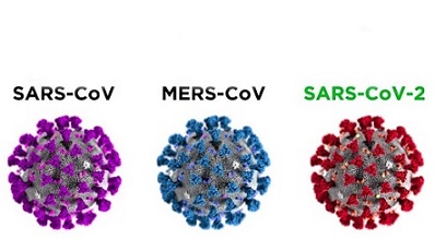
SARS-CoV-2 Is Different From All Other Betacoronaviruses In That It Only Partially Activates The IRE1α/XBP1 Endoplasmic Reticulum Stress Pathway
Source: Medical News -SARS-CoV-2 Research Oct 03, 2022 3 years, 2 months, 3 weeks, 5 days, 18 hours, 39 minutes ago
A new study led by researchers from the University of Chicago that involved medical scientists from the University of Pennsylvania has found that the SARS-CoV-2 coronavirus is different from all other known Betacoronaviruses in that it only partially activates the IRE1α/XBP1 endoplasmic reticulum stress pathway.

Corresponding author, Dr Susan R. Weiss from the Department of Microbiology, University of Pennsylvania told Thailand
Medical News, “SARS-CoV-2 only partially activates IRE1α, promoting its kinase activity but not RNase activity. Based on IRE1α-dependent gene expression changes during infection, we propose that SARS-CoV-2 prevents IRE1α RNase activation as a strategy to limit detection by the host immune system.”
The IRE1α-XBP1 pathway is the most conserved branch of the unfolded protein response pathways, which are activated during endoplasmic reticulum (ER) stress caused by the accumulation of unfolded/misfolded proteins in the ER lumen. The IRE1α-XBP1 pathway plays a critical role in various cancers especially in melanomas.
The SARS-CoV-2 coronavirus has killed over 6.55 million individuals worldwide and infected over 623 million people worldwide according to reported figures. (In reality the actual figures could be 5 to 6-fold!).
With the continuous emergence of new variants and coming Winter surges that are expected to be catastrophic, it is critical to unravel the molecular mechanisms that allow SARS-CoV-2 and other coronaviruses to infect and overtake the host machinery of human cells.
Typically, coronavirus replication triggers endoplasmic reticulum (ER) stress and activation of the unfolded protein response (UPR), a key host cell pathway widely believed to be essential for viral replication.
The study team examined the master UPR sensor IRE1α kinase/RNase and its downstream transcription factor effector XBP1s, which is processed through an IRE1α-mediated mRNA splicing event, in human lung-derived cells infected with betacoronaviruses.
The study findings showed that human respiratory coronavirus OC43 (HCoV-OC43), Middle East respiratory syndrome coronavirus (MERS-CoV), and murine coronavirus (MHV) all induce ER stress and strongly trigger the kinase and RNase activities of IRE1α as well as XBP1 splicing.
However, in contrast….the SARS-CoV-2 coronavirus only partially activates IRE1α through autophosphorylation, but its RNase activity fails to splice XBP1.
Furthermore, while IRE1α was dispensable for replication in human cells for all coronaviruses tested, it was required for maximal expression of genes associated with several key cellular functions, including the interferon signaling pathway, during SARS-CoV-2 infection.
The study findings suggest that SARS-CoV-2 actively inhibits the RNase of autophosphorylated IRE1α, perhaps as a strategy to eliminate detection by the host immune system.
The study findings were published in the peer reviewed journal: mBio.
https://journals.asm.org/doi/10.1128/mbio.02415-22
r />
The SARS-CoV-2 virus is the third lethal respiratory coronavirus, after MERS-CoV and SARS-CoV, to emerge this century, causing a major health crisis. The other common coronaviruses such as HCoV-OC43 causes less severe respiratory disease. Hence, it is imperative to understand the similarities and differences among these viruses in how each interacts with host cells.
The study team focused here on the inositol-requiring enzyme 1α (IRE1α) pathway; part of the host unfolded protein response to virus-induced stress. Our study findings showed that while MERS-CoV and HCoV-OC43 fully activate the IRE1α kinase and RNase activities, SARS-CoV-2 only partially activates IRE1α, promoting its kinase activity but not RNase activity. Based on IRE1α-dependent gene expression changes during infection, we propose that SARS-CoV-2 prevents IRE1α RNase activation as a strategy to limit detection by the host immune system.
In order to better understand the host epithelial response to coronavirus infection, the study team systematically compared the activation of the IRE1α/XBP1 pathway of the UPR during infection with betacoronaviruses in lung-derived A549 and Calu-3 cells lines and iPSC-derived AT2 cells.
The study team employed three human viruses, each from a different betacoronavirus subgenus, OC43 (embeco), SARS-CoV-2 (sarbeco) and MERS-CoV (merbeco), and included the murine coronavirus MHV, a model embecovirus.
The study team found a striking difference between the host response to SARS-CoV-2 and the other three viruses.
It was found that OC43, MHV, and MERS-CoV all activated the canonical IRE1α/XBP1 pathway in both A549 and Calu-3 cell lines as evidenced by phosphorylation of IRE1α, XBP1 mRNA splicing and induction of DNAJB9, a transcriptional target of XBP1s.
Furthermore, MERS-CoV was observed to induce IRE1α/XBP1 activation in iAT2 cells.
Interestingly, while SARS-CoV-2 also promoted autophosphorylation of IRE1α, there was no evidence of XBP1s, indicating that the pathway was only partially activated and suggesting that the IRE1α kinase was active while the XBP1 splicing RNase activity was not.
The differential splicing of XBP1 mRNA during SARS-CoV-2 and MERS-CoV infection was also observed in iPSC-derived AT2 cells, confirming the results in a more physiologically relevant system.
The complete difference among these viruses is surprising, as all of them encode highly conserved replicase and structural proteins that promote ER membrane rearrangements and challenge the ER folding capacity, respectively.
The study team had originally hypothesized that these conserved genes would induce similar stress on the ER and lead to UPR activation.
However, the study findings suggest that that SARS-CoV-2 actively prevents XBP1 splicing.
Consistent with this idea, a recombinant SARS-CoV lacking the E protein (rSARS-CoV-ΔE) was reported to induce more XBP1 splicing as well as induction of UPR genes compared to parental wild-type virus.
In order to investigate the importance of IRE1α for coronavirus replication, the study team evaluated replication of each of the betacoronaviruses in IRE1α KO A549 cells compared to parental wild-type cells.
In contrast to influenza, all of the betacoronaviruses examined were able to replicate efficiently in the absence of IRE1α signaling, consistent with a previous report of the gammacoronavirus IBV.
https://pubmed.ncbi.nlm.nih.gov/25142592/
Although the study team did observe a decrease in OC43 and SARS-CoV-2 nucleocapsid expression following KIRA8 treatment, the similar levels of replication of all the viruses in IRE1α KO cells and parental cells suggest that this is due to off-target effects of KIRA8 rather than IRE1α inhibition limiting virus replication.
Hence, this raises interesting possibilities for the role of IRE1α during coronavirus infection.
It was already previously known that IRE1α can produce both cytoprotective (through XBP1s) and destructive responses (via RIDD and JNK/p38 signaling) depending on the extent of the encountered stress.
It seems likely that coronavirus infection would induce extensive and prolonged ER stress, which may push IRE1α beyond the initial pro-recovery responses and toward a pro-apoptotic response.
The study data did indeed reveal that, at least with MERS-CoV and SARS-CoV-2 infection, IRE1α phosphorylation is readily detectable by 24 hpi and remains steady throughout the course of infection.
Also, unlike what has been observed with chemically induced ER stress, IRE1α phosphorylation does not appear to attenuate at any point during coronavirus infection, again suggesting a hyperactive and destructive outcome.
Destruction of cells, in particular, AT2 cells in the lung, may contribute to pathogenesis during coronavirus infection. However, SARS-CoV-2 appears to limit the downstream consequences of IRE1α activation, most notably, XBP1 splicing via its RNase activity, and thus may be protected from this destructive phenotype. MERS-CoV may induce apoptosis redundantly in the UPR, as it has been reported that MERS-CoV induces and benefits from apoptosis mediated by the PERK arm of the UPR.
In order to further probe the impact of IRE1α signaling on host gene expression following coronavirus infection, the study team performed RNA-seq analysis of sg control or IRE1α knockout A549-ACE2 cells infected with either SARS-CoV-2 or OC43.
It was found that IRE1α deletion significantly reduced the expression of genes downstream of XBP1s during OC43 infection, as expected, with otherwise only modest changes in overall gene expression.
However, genetic ablation of IRE1α significantly impacted host gene expression in SARS-CoV-2-infected A549 cells. The two most dramatic effects that appear to be specific to SARS-CoV-2 relate to chromatin organization and protein folding and transport. Effects on mRNA metabolism and processing are also observed for SARS-CoV-2 and, more modestly, for OC43. Finally, protein translation is downregulated in both OC43 and SARS-CoV-2-infected cells but, in the latter case, occurs primarily upon loss of IRE1α.
The study findings suggest that IRE1α plays a key role in mediating changes in host cell gene transcription and protein production caused by SARS-CoV-2.
The study team found here that deletion of IRE1α blunted the induction of some but not all ISGs by SARS-CoV-2 infection.
In contrast, OC43 was not observed to induce significant levels of IFN or ISG mRNAs in either WT or IRE1α KO cells.
Importantly, the mechanism by which loss of IRE1α activity during SARS-CoV-2 infection dampens the induction of interferon signaling remains to be determined.
It has been reported that the UPR can precede and prime innate immune signaling in flavivirus-infected cells.
https://pubmed.ncbi.nlm.nih.gov/31467282/
XBP1s has been found upstream of IFNα and IFNβ transcription and may work through binding upstream cis-acting enhancer elements.
https://pubmed.ncbi.nlm.nih.gov/28408069/
https://pubmed.ncbi.nlm.nih.gov/20660350/
Also, XBP1s can directly bind and transcriptionally activate interleukin-6 (IL-6), tumor necrosis factor α (TNF-α), and other inflammatory cytokines.
https://pubmed.ncbi.nlm.nih.gov/29556237/
The study team said that it is possible that a low level of background XBP1 splicing may occur during SARS-CoV-2 infection, which could contribute to these responses. Independent of its RNase activity, the autophosphorylated cytoplasmic domain of IRE1α can oligomerize and serve as a scaffold that recruits TRAF2, JNK, ASK, Nck, and other molecules that can lead to varied signaling outputs.
Hence, the ability of SARS-CoV-2 coronavirus to prevent full IRE1α activation might dampen inflammatory signaling and prevent detection and elimination by the immune system in an intact organism.
It is important to note that the diminution of ISG expression in the absence of IRE1α is variable among ISGs, and SARS-CoV-2 still induces IFN and IFN signaling to a greater extent than OC43 in IRE1α KO cells.
The study team speculates that SARS-CoV-2 has adapted to tolerate a low level of IFN signaling as well as protein kinase R (PKR) and oligoadenylate RNase L (OAS/RNase L) activation, and the reduced ISG expression in the absence of IRE1α does not have enough of an effect to promote increased replication. This is consistent with our finding that knockout of mitochondrial antiviral signaling protein (MAVS) from A549 cells, resulting in minimal IFN expression and ISG signaling, does not promote increased SARS-CoV-2 replication.
https://pubmed.ncbi.nlm.nih.gov/33811184/
Hence, the significance of IRE1α-dependent IFN signaling is not clear and will be a subject of future investigation.
The study findings show that despite the lack of apparent virus replication defects with IRE1α deficiency, further characterization of the repertoire of betacoronavirus-induced IRE1α signaling is warranted, including contributions to cytokine production, apoptosis, and proinflammatory responses.
Although the study team initially investigated this pathway from the perspective of the impact on virus replication, future studies should examine effects of IRE1α activation on the host, including inflammation and cell death through the JNK and p38 mitogen-activated protein kinase (MAPK) signaling scaffolded by IRE1α and/or RIDD, as a consequence of prolonged IRE1α activation.
Such responses could be particularly important in AT2 cells, which must rely on the UPR to maintain proteostasis in the face of the challenge from the biosynthesis and secretion of surfactant proteins.
Any dysregulation of these responses by coronavirus infection could promote AT2 cell reprogramming, epithelial apoptosis, alteration of surfactant components in alveoli, and the rampant inflammation associated with severe coronavirus infection.
The study team recently reported that SARS-CoV-2 and MERS-CoV also diverge in their activation and antagonism of the dsRNA-induced host cell innate immune responses, another early innate response to viruses.
https://pubmed.ncbi.nlm.nih.gov/33811184/
Interestingly, while MERS-CoV actively antagonizes type I and type III interferon production and signaling, the oligoadenylate RNase L (OAS/RNase L) system and the PKR pathway, SARS-CoV-2 activates OAS/RNase L and PKR and induces a low level of IFN and ISG expression in A549 and Calu-3 respiratory tract-derived cells.
https://pubmed.ncbi.nlm.nih.gov/30914508/
It was observed that OC43 infection did not lead to the induction of IFN or ISGs, and it was already shown previously that OC43-encoded accessory protein NS2 antagonizes activation of the OAS/RNase L pathway.
https://pubmed.ncbi.nlm.nih.gov/28003490/
Activation of these pathways during MERS-CoV mutant infection significantly reduces virus replication, whereas SARS-CoV-2 can tolerate the innate responses activated during infection.
The study team commented, “Considering the differences we have observed between betacoronaviruses with innate immune responses and now IRE1α activation and signaling, it is striking that MERS-CoV and SARS-CoV-2 are reciprocal in what they activate and antagonize. To optimize replication, coronaviruses must likely strike a balance in the cellular responses they antagonize, tolerate, or benefit from. Supporting this, our data suggest that IRE1α influences ISG induction during infection. It is intriguing to consider if MERS-CoV tolerates this by antagonizing IFN and ISG induction, while SARS-CoV-2 instead limits IRE1α activity. Future studies should examine the synergy between innate immune responses and the UPR during coronavirus infection and how perturbations on one side may change viral replicative capacity, tropism, and spread. Understanding how signals from each one of these pathways are integrated into viral replication and cell fate decisions during coronavirus infection may illuminate new therapeutic strategies for combating emerging betacoronaviruses.”
For the latest SARS-CoV-2 Research, keep on logging to Thailand Medical News.
