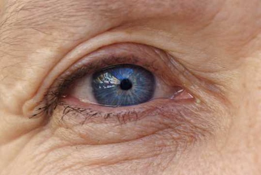
Scientist Discover How A Protein Called Vitronectin Causes Macular Degeneration, An Eye Disease That Affects More Than 200 Million People!
Source: Medical News - Vitronectin - Macular Degeneration Sep 07, 2022 2 years, 7 months, 2 weeks, 5 days, 12 hours, 18 minutes ago
Researchers from the independent biomedical research institute located in California called Sanford Burnham Prebys have identified the mechanism by which the blood protein called Vitronectin causes macular degeneration.

Macular degeneration is as common eye disorder among people over 50. It causes blurred or reduced central vision and many normally end up being blinded. It is estimated that almost 200 people globally are infected with the disease.
The study team had previously identified vitronectin as the blood protein responsible of the formation of deposits in dry age-related macular degeneration.
https://www.pnas.org/doi/10.1073/pnas.2007699117
Senior author of the study, Dr Francesca Marassi, director of the Cancer, Molecules and Structures Program at Sanford Burnham Prebys Medical Discovery Institute told Thailand
Medical News, “Our previous findings suggest that vitronectin, which is shaped like a sticky propeller, orchestrates the formation of the spherical deposits that accumulate and cause dry age-related macular degeneration (AMD). With this information, we can look for drugs that prevent the deposits from forming and help people retain their sight for as long as possible.”
It was reported that more than 11 million individuals in the United States alone have AMD, of which the dry variation accounts for 80 to 90 percent of cases. While the progression of the disease can be slowed with dietary and lifestyle changes, no pharmaceutical treatment option currently exists.
It was found that dry AMD is caused by the progressive accumulation of drusen, pebble-like deposits at the back of the eye, which results in blurry vision and eventually blindness for many!
Although scientists knew these deposits contain cholesterol, lipids, proteins such as vitronectin and a mineralized form of calcium phosphate called hydroxyapatite (the material that makes up bones and teeth), how these deposits form was unknown.
In that study, Dr Marassi and colleagues used the structure of vitronectin and various biophysical tools to prove that the propeller top tightly clasps calcium and hydroxyapatite. According to the team, the findings suggest a mechanism by which vitronectin drives the formation of the abnormal deposits and reveals how this process might be interrupted.
Dr Marassi added, “We know that these deposits have a cholesterol-rich lipid core that is surrounded by a shell of hydroxyapatite and a final topcoat of vitronectin. Our study suggests that vitronectin brings all these pieces together in one place to build up this complex assembly. With this information, we can start to figure out how to disrupt these interactions and break up this deposit.”
The study team has been already working with scientists at the Institute’s Conrad Prebys Center for Chemical Genomics to identify vitronectin-targeting compounds that can stop drusen from forming. This drug candidate would hold promise as a treatment to slow the progression of dry AMD and potentially other plaque-related conditions, such as Alzheimer’s where vitronectin is known to be a major component of amyloid plaques.
In the n
ew study, the study team managed to discover the molecular secrets of macular degeneration, and the detailed mechanism by which vitronectin contributes to macular degeneration.
The study team stated in their study abstract that the adaptability of proteins to their work environments is fundamental for cellular life.
The study team found how the hemopexin-like (HX) domain of the multifunctional blood glycoprotein vitronectin (Vn) binds Ca2+ to adapt to excursions of temperature and shear stress.
Utilizing X-ray crystallography, molecular dynamics (MD) simulations, nuclear magnetic resonance (NMR) and differential scanning fluorimetry (DSF), the study team described how Ca2+ and its flexible hydration shell enable the protein to perform conformational changes that relay beyond the Ca2+ binding site, and alter the number of polar contacts to enhance conformational stability.
By means of mutagenesis, the study team identified key residues that cooperate with Ca2+ to promote protein stability, and the researchers showed that Ca2+ association confers protection against shear stress, a property that is advantageous for proteins that circulate in the vasculature, like Vn.
The study findings were published in the peer reviewed Biophysical Journal.
https://www.cell.com/biophysj/fulltext/S0006-3495(22)00726-3
This is the first study findings to describe in detail the flexible structure of the key blood protein ie Vitronectin involved in macular degeneration and other age-related diseases, such as Alzheimer’s and atherosclerosis.
Dr Marassi told Thailand
Medical News, “Proteins in the blood are under constant and changing pressure because of the different ways blood flows throughout the body. For example, blood flows more slowly through small blood vessels in the eyes compared to larger arteries around the heart. Blood proteins need to be able to respond to these changes, and this study gives us fundamental truths about how they adapt to their environment, which is critical to targeting those proteins for future treatments.”
Although there are hundreds of proteins in our blood, the study team focused on vitronectin, one of the most abundant as a result of the previous study findings mentioned above.
Most importantly, in addition to circulating in high concentrations in the blood, vitronectin is found in the scaffolding between cells and is also an important component of cholesterol.
Dr Marassi explained, “This protein is an important target for macular degeneration because it accumulates in the back of the eye, causing vision loss. Similar deposits appear in the brain in Alzheimer’s disease and in the arteries in atherosclerosis. We want to understand why this happens and leverage this knowledge to develop new treatments.”
The study team was interested in learning how the protein changes its structure at different temperatures and under different levels of pressure, approximating what happens in the human body.
Dr Marassi added, “Determining the structure of a protein is the most important part of determining its function.”
Utilizing detailed biochemical analysis, the study team found that the protein can subtly change its shape under pressure. These changes cause it to bond more easily to calcium ions in the blood, which the stud team suggests leads to the buildup of calcified plaque deposits characteristic of macular degeneration and other age-related diseases.
Dr Marassi further explained, “It’s a very subtle rearrangement of the molecular structure, but it has a big impact on how the protein functions. The more we learn about the protein on a structural and mechanistic level, the better chance we have of successfully targeting it with treatments.”
Most significantly, these structural insights will streamline the development of treatments for macular degeneration because it will allow researchers and their partners in the biotech industry to custom-design antibodies that selectively block the protein’s calcium binding without disrupting its other important functions in the body.
Dr Marassi added, “It will take some time to convert it into a clinical treatment, but we hope to have a working antibody as a potential treatment in a few years’ time. And since this protein is so abundant in the blood, there may be other exciting applications for this new knowledge that we don’t even know about yet.”
For more on
Vitronectin and Macular Degeneration, keep on logging to Thailand
Medical News.
