Nikhil Prasad Fact checked by:Thailand Medical News Team Apr 28, 2024 1 year, 8 months, 3 days, 44 minutes ago
Cancer News: In the realm of cancer treatment, personalized medicine has emerged as a groundbreaking approach, tailoring therapies to the specific needs of individual patients based on their unique cancer type and characteristics. A recent pioneering study led by Professor Josep Sulé-Suso at Keele University that is covered in this
Cancer News report, has demonstrated a new method for identifying single cancer cells within blood samples, marking a significant advancement in cancer diagnosis and treatment strategies.
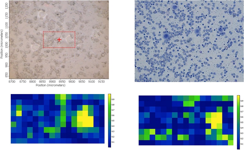
 New Method Of Identifying Single Cancer Cells In Blood. Brightfield image of a cytospun sample containing PBMC and individual CALU-1 cancer cells (A). The same image following Giemsa staining (B). FTIR spectra false colour intensity maps of the spatial rectangular region showed in A for the lipid and Amide A region (C) and the fingerprint region between 1350 cm-1 and 1800 cm-1 (D).
The Significance of Liquid Biopsy
Liquid biopsy has garnered global interest as a promising avenue to enhance cancer management. It involves analyzing various components in bodily fluids, such as circulating tumor DNA, RNA, exosomes, and circulating tumor cells (CTCs). These techniques offer diverse applications, including early cancer detection, treatment response assessment, and personalized therapy selection based on tumor subtypes.
CTCs are cancer cells present in peripheral blood, originating from primary tumors or metastases. Detecting and analyzing CTCs play a crucial role in understanding tumor progression, metastasis, and treatment response. However, existing methods for isolating and identifying CTCs face challenges in robustness and accuracy.
Challenges in CTC Identification
Current approaches to isolate CTCs rely on antigen expression or physical properties, leading to limitations in capturing all types of CTCs effectively. Positive selection methods based on cancer markers may overlook cells lacking specific antigens, while negative selection methods against blood cell antigens can be cumbersome and costly. Additionally, the phenomenon of Epithelial–Mesenchymal Transition (EMT) further complicates CTC identification, as it alters cell properties, making traditional isolation methods less reliable.
FTIR Microspectroscopy: A Game-Changing Technology
Fourier Transform Infrared (FTIR) microspectroscopy emerges as a potential game-changer in CTC identification. This technique leverages the biochemical composition of cells, utilizing infrared light to differentiate between cancerous and non-cancerous cells. Unlike traditional methods, FTIR microspectroscopy is not dependent on specific markers or physical properties, making it a versatile and potentially robust tool for CTC detection.
Feasibility Study and Experimental Insights
In a groundbreaking feasibility study, researchers utilized FTIR microspectroscopy to identify individual lun
g cancer cells within blood samples. The study focused on lung cancer cells mixed with peripheral blood mononuclear cells (PBMCs), showcasing the capability of FTIR microspectroscopy to distinguish between cancerous and non-cancerous cells based on their biochemical signatures.
Biochemical Signatures and FTIR Microspectroscopy
The study highlighted specific regions within the infrared spectra, such as the lipid and Amide A regions, as crucial for cancer cell identification. These regions provide insights into the lipid content, membrane composition, and protein structure of cells, which differ significantly between cancerous and non-cancerous cells. The use of FTIR microspectroscopy facilitated the accurate identification of single cancer cells amidst complex cellular environments, offering a promising avenue for liquid biopsy applications.
Professor Sule-Suso commented, "Identifying cancer cells in blood using this new method could be a game-changer in the management of patients with cancer."
Translating Research to Clinical Applications
The successful demonstration of FTIR microspectroscopy in identifying single cancer cells paves the way for its integration into clinical practice. Overcoming challenges such as substrate compatibility and data processing speed are key priorities for advancing this technology towards widespread clinical adoption. Integration with existing pathology techniques, such as staining and immunohistochemistry, further enhances its utility in cancer diagnosis and characterization.
Professor Kamaraj Karunanithi, Director of Research and Innovation at University Hospitals of North Midlands NHS Trust (UHNM) that was also involved in the research and development commented, "The UHNM Research and Innovation Department is thrilled to back research targeting the early detection of cancer cells in the bloodstream. Detecting cancer spread early is vital for improving patient care. This study has the potential to establish new standards in treating cancer at various stages. The UHNM research strategy not only backs delivery of national and international research but also concentrates on identifying and developing local research in collaboration with our local universities."
Future Directions and Impact
Ongoing research aims to expand the scope of FTIR microspectroscopy to encompass a wide range of cancer types, validating its effectiveness across diverse patient populations. Improvements in instrumentation and algorithm development will streamline the process, making FTIR microspectroscopy a valuable tool for routine cancer screening, treatment monitoring, and personalized therapy decisions.
Conclusion
The convergence of advanced technologies like FTIR microspectroscopy with the evolving landscape of personalized medicine heralds a new era in cancer diagnosis and treatment. By enabling the precise identification of single cancer cells in blood samples, FTIR microspectroscopy empowers clinicians with actionable insights for tailored patient care. As research progresses and methodologies refine, the promise of liquid biopsy and FTIR microspectroscopy in revolutionizing cancer management becomes increasingly tangible.
The study findings were published in the peer reviewed journal: PLOS One
https://journals.plos.org/plosone/article?id=10.1371/journal.pone.0289824
For the
Cancer News, keep on logging to Thailand Medical News.
