Scientists Discover Shocking Pulmonary Microvascular Abnormalities Lingering Months After Non-Severe COVID-19 Infections!
COVID-19 News - Pulmonary Microvascular Abnormalities - Months After Non-Severe COVID-19 Infections Jun 18, 2023 2 years, 6 months, 4 weeks, 18 hours, 25 minutes ago
Harvard Study Using Cutting-Edge Imaging Technology Reveals That Many With Only Mild COVID-19 Could Suffer From Pulmonary Vascular Dysfunction!
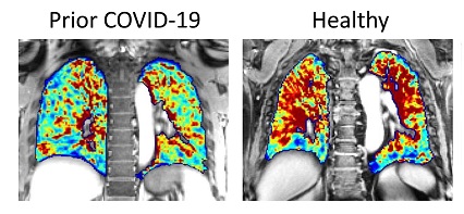
COVID-19 News: In a new study conducted by Harvard Medical School and Massachusetts General Hospital, scientists have made a stunning discovery regarding the long-term effects of COVID-19 on the human body. Using a revolutionary imaging technique called dynamic contrast-enhanced magnetic resonance imaging (DCE-MRI), researchers have uncovered pulmonary microvascular abnormalities in individuals who experienced non-severe SARS-CoV-2 infections several months earlier. These findings shed new light on the vascular complications caused by the virus and raise questions about its long-term impact on survivors.

While studies and
COVID-19 News reports have already showed that SARS-CoV-2-2 could cause microvascular damage, whether such damage is long lasting has never been fully addressed especially concerning the lungs.
https://www.thailandmedical.news/news/scientists-validate-that-covid-19-causes-microvascular-alteration-in-study-using-nailfold-capillaroscopy
https://www.thailandmedical.news/news/long-covid-news-microvasculature-alterations,-distinct-transcriptomic-signatures-in-skeletal-muscles-and-macrophage-infiltration-found-in-long-covid
https://www.thailandmedical.news/news/must-read-covid-19-is-nothing-but-a-microvascular-and-endothelial-disease-that-kills-fast-or-slowly-depending-on-the-human-host-health-status
https://www.thailandmedical.news/news/long-covid-news-study-shows-majority-of-young-healthy-adults-will-exhibit-peripheral-macrovascular-and-microvascular-complications-following-covid-19
Studies have also showed microvascular damage occurring in organs like the brain and heart due to SARS-CoV-2 infections.
https://www.thailandmedical.news/news/u-s-nih-s-and-u-s-army-s-neurocovid-study-shows-that-sars-cov-2-infections-causes-serious-damage-to-microvessels-in-the-brain
pread-prevalence-of-inflammatory-microvasculopathy-in-brains-of-infected">https://www.thailandmedical.news/news/breaking-study-alarmingly-reveals-that-sars-cov-2-infections-causes-widespread-prevalence-of-inflammatory-microvasculopathy-in-brains-of-infected
https://www.thailandmedical.news/news/breaking-german-scientists-discover-that-sars-cov-2-main-protease-mpro-causes-microvascular-brain-pathology
https://www.thailandmedical.news/news/murine-study-reveals-yet-another-way-that-sars-cov-2-causes-heart-damage-targeting-cardiac-pericytes,-causing-heart-s-microvasculature-damage
However, one German study did show vascular damage up to 18 months after COVID-19 infection!
https://www.thailandmedical.news/news/breaking-german-study-shows-persistent-capillary-rarefication-a-reduction-in-vascular-density-even-18-months-after-covid-19-infection
Unmasking the Vascular Effects of COVID-19
COVID-19 has been known to wreak havoc on the body, primarily affecting the respiratory system. However, the extent of vascular endothelial dysfunction in non-severe cases has remained elusive until now. The team of scientists behind this study had previously used DCE-MRI to detect microvascular differences in patients with idiopathic pulmonary fibrosis. They hypothesized that this innovative imaging technique could also uncover similar microvascular changes in individuals recovering from symptomatic COVID-19.
Study Design and Findings
The study included participants who had tested positive for SARS-CoV-2 within the past 3–12 months and healthy volunteers without known lung disease. DCE-MRI was performed using state-of-the-art equipment, capturing dynamic images of the lungs before, during, and after the injection of a contrast agent. By analyzing the signal intensity versus time curve, the researchers gained valuable insights into microvascular perfusion and extravascular extracellular space.
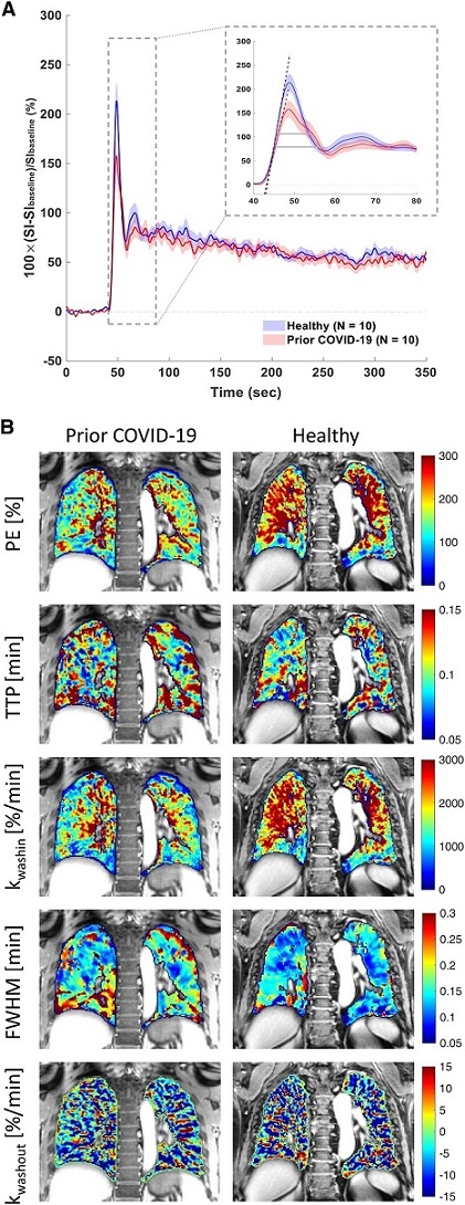
Ten participants with prior COVID-19 and ten healthy volunteers underwent DCE-MRI. The results were astonishing. The signal versus time curves of individuals who had recovered from COVID-19 exhibited reduced pulmonary microvascular perfusion compared to those of the healthy volunteers. Key findings included slower contrast arrival rates, broader peaks, and a trend towards lower peak enhancement magnitude in the COVID-19 group. Surprisingly, the rate of contrast washout did not differ significantly between the two groups, suggesting a lack of residual tissue fibrosis or edema.
 Dynamic contrast-enhanced magnetic resonance imaging of prior coronavirus disease participants versus healthy volunteers. (A) Magnetic resonance imaging signal intensity (SI) versus time curves of the lung parenchyma from healthy volunteers (n = 10) and participants with prior coronavirus disease (COVID-19) (n = 10) computed as percentage of SI change relative to the lung signal before gadolinium administration. The group-averaged dynamic curves from posterior coronal regions of interest are shown. Shaded area indicates mean ± 1 SEM. The inset shows the zoom-in of the first-pass peaks, with dashed lines indicating the upslopes and gray horizontal bars indicating the full width at half maximum (FWHM) of the first-pass peaks. (B) Representative parametric maps of peak enhancement, TTP, kwashin, FWHM, and kwashout from a posterior coronal region of interest of a participant who had prior COVID-19 and who did not require hospitalization and a healthy volunteer. kwashin = rate of contrast washin; kwashout = late-phase washout slope between 60 seconds postinjection and the last acquisition; PE = peak enhancement; TTP = time to peak enhancement.
Implications and Future Research
These findings have significant implications for understanding the long-term effects of COVID-19 on the human body. The study demonstrated that DCE-MRI is a highly sensitive tool for detecting pulmonary microvascular abnormalities, even in individuals with normal pulmonary function. This suggests that the impact of COVID-19 on the vasculature may be more widespread and enduring than previously thought.
While this study's sample size was small, the results pave the way for further research into the connection between DCE-MRI measurements and other clinical parameters, such as chest computed tomography (CT) findings, pulmonary function abnormalities, and persistent respiratory symptoms. Additionally, researchers plan to investigate the specific mechanisms behind these microvascular abnormalities, including residual microvascular thrombosis or vascular remodeling.
A Call to Action
These groundbreaking findings highlight the urgent need for continued research and monitoring of COVID-19 survivors, even those who experienced mild or non-severe infections. The lingering effects on the pulmonary vasculature could have profound implications for future treatment strategies and the development of targeted therapies for acute and post-acute stages of the disease.
As the scientific community works tirelessly to unravel the mysteries of COVID-19, studies like this one bring us closer to understanding the virus's full impact. It is imperative that these findings reach a wide audience, igniting discussions among medical professionals, researchers, and the general public. By sharing this groundbreaking study, we can raise awareness about the long-term consequences of COVID-19 and inspire further research to improve patient care and treatment outcomes.
Disclaimer
The findings presented in this study are preliminary, and further research is necessary to fully comprehend the implications of pulmonary microvascular abnormalities in COVID-19 survivors.
The study findings were published in the peer reviewed American Journal of Respiratory and Critical Care Medicine.
https://www.atsjournals.org/doi/full/10.1164/rccm.202210-1884LE
For the latest
COVID-19 News, keep on logging to Thailand Medical News.

