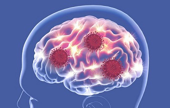
Study Confirms That Even In Asymptomatic To Mild Cases Of Infections, SARS-CoV-2 Leaves A Mark On The Brain!
Source: SARS-CoV-2-Brain Sep 28, 2021 4 years, 4 months, 2 weeks, 1 day, 9 hours, 11 minutes ago
SARS-CoV-2-Brain: A new preliminary but large-scale study led researchers from the Department of Clinical Neurosciences of the University of Oxford-UK, along with scientist from National Institutes of Mental Health, National Institutes of Health-USA and the Department of Brain Sciences, Imperial College-UK investigating brain changes in individuals who had experienced COVID-19 drew a great deal of attention within the neuroscience community.

The study findings revealed that even in asymptomatic to mild cases of infections, the SARS-CoV-2 coronavirus leaves a mark on the human host’s brain!
The research team studied the possible brain changes associated with the coronavirus infection using multimodal MRI data from 785 adult participants (aged 51–81) from the UK Biobank COVID-19 re-imaging study, including 401 adult participants who tested positive for SARS-CoV-2 infection between their two scans. The team used structural, diffusion and functional brain scans from before and after infection, to compare longitudinal changes between these 401 SARS-CoV-2 cases and 384 controls who had either tested negative to rapid antibody testing or had no COVID-19 medical and public health record, and who were matched to the cases for age, sex, ethnicity and interval between scans. The controls and cases did not differ in blood pressure, body mass index, diabetes diagnosis, smoking, alcohol consumption, or socio-economic status.
Utilizing both hypothesis-driven and exploratory approaches, with false discovery rate multiple comparison correction, we identified respectively 68 and 67 significant longitudinal effects associated with SARS-CoV-2 infection in the brain, including, on average: (i) a more pronounced reduction in grey matter thickness and contrast in the lateral orbitofrontal cortex (min P=1.7×10⁻⁴, r=-0.14) and parahippocampal gyrus (min P=2.7×10⁻⁴, r=-0.13), (ii) a relative increase of diffusion indices, a marker of tissue damage, in the regions of the brain functionally-connected to the piriform cortex, anterior olfactory nucleus and olfactory tubercle (min P=2.2×10⁻⁵, r=0.16), and (iii) greater reduction in global measures of brain size and increase in cerebrospinal fluid volume suggesting an additional diffuse atrophy in the infected participants (min P=4.0×10⁻⁶, r=-0.17).
When looking over the entire cortical surface, these grey matter thickness results covered the parahippocampal gyrus and the lateral orbitofrontal cortex, and extended to the anterior insula and anterior cingulate cortex, supramarginal gyrus and temporal pole. The increase of a diffusion index (mean diffusivity) meanwhile could be seen voxel-wise mainly in the medial and lateral orbitofrontal cortex, the anterior insula, the anterior cingulate cortex and the amygdala.
The study findings were not altered after excluding cases who had been hospitalized. The team further compared hospitalized (n=15) and non-hospitalized (n=386) infected participants, resulting in similar findings to the larger cases vs control group comparison, with, in addition, a marked reduction of grey matter thickness in fronto-parietal and temporal regions (all FDR-significant, min P=4.0×10⁻⁶). The 401 SARS-CoV-2 infected participants also showed larger cognitive decline between the two timepoints in the Trail Making Test compared with the controls (both FDR-significant, min P=1.0×10⁻⁴, r=0.17; and still FDR-significant after excluding the hospitalized p
atients: min P=1.0×10⁻⁴, r=0.17), with the duration taken to complete the alphanumeric trail correlating post hoc with the cognitive and olfactory-related crus II of the cerebellum (FDR-significant, P=2.0×10⁻³, r=-0.19), which was also found significantly atrophic in the SARS-CoV-2 participants (FDR-significant, P=6.1×10⁻⁵, r=-0.14).
The study findings thus relate to longitudinal abnormalities in limbic cortical areas with direct neuronal connectivity to the primary olfactory system. Unlike in post hoc cross-sectional studies, the availability of pre- infection imaging data mitigates to some extent the issue of pre-existing risk factors or clinical conditions being misinterpreted as disease effects.
The
SARS-CoV-2-Brain study team was therefore able to demonstrate that the regions of the brain that showed longitudinal differences post-infection did not already show any difference between (future) cases and controls in their initial, pre-infection scans.
These brain imaging results may be the in vivo hallmarks of a degenerative spread of the disease or of the virus itself via olfactory pathways (a possible entry point of the virus to the central nervous system being via the olfactory mucosa), or of neuroinflammatory events due to the infection, or of the loss of sensory input due to anosmia. Whether this deleterious impact can be partially reversed, for instance after improvement of the hyposmic symptoms, or whether these are effects that will persist in the long term, remains to be investigated with additional follow up.
The study findings were published on a preprint server and are currently being peer reviewed.
https://www.medrxiv.org/content/10.1101/2021.06.11.21258690v3
Almost 21 months into the pandemic, researchers have been steadily gathering new and important insights into the effects of COVID-19 on the body and brain. These findings are raising concerns about the long-term impacts that the coronavirus might have on biological processes such as aging.
In the study, the research team analyzed the brain imaging data and then brought back those who had been diagnosed with COVID-19 for additional brain scans. The study team compared individuals who had experienced COVID-19 to participants who had not, carefully matching the groups based on age, sex, baseline test date and study location, as well as common risk factors for disease, such as health variables and socioeconomic status.
The study team found marked differences in gray matter which is made up of the cell bodies of neurons that process information in the brain, between those who had been infected with COVID-19 and those who had not.
Shockingly, the thickness of the gray matter tissue in brain regions known as the frontal and temporal lobes was reduced in the COVID-19 group, differing from the typical patterns seen in the group that hadn't experienced COVID-19.
In the general population, it is normal to see some change in gray matter volume or thickness over time as people age, but the changes were larger than normal in those who had been infected with COVID-19.
Importantly when the study team separated the individuals who had severe enough illness to require hospitalization, the results were the same as for those who had experienced milder COVID-19 or were asymptomatic.
Significantly that showed that as long as people were infected with the SARS-CoV-2 coronavirus irrespective as to whether they were asymptomatic or had only mild symptoms or were hospitalized due to disease severity, they showed a loss of brain volume!
The study team also investigated changes in performance on cognitive tasks and found that those who had contracted COVID-19 were slower in processing information, relative to those who had not.
Although one has to be careful interpreting these findings as they await formal peer review, the large sample, pre- and post-illness data in the same people and careful matching with people who had not had COVID-19 have made this preliminary work particularly valuable.
In the initial stages of the pandemic, one of the most common reports from those infected with COVID-19 was the loss of sense of taste and smell.
Interestingly, the brain regions that the study team found to be impacted by COVID-19 are all linked to the olfactory bulb, a structure near the front of the brain that passes signals about smells from the nose to other brain regions.
It should be noted that the olfactory bulb has connections to regions of the temporal lobe. We often talk about the temporal lobe in the context of aging and Alzheimer's disease because it is where the hippocampus is located. The hippocampus is likely to play a key role in aging, given its involvement in memory and cognitive processes.
It should be noted that the sense of smell is also important to Alzheimer's research, as some data has suggested that those at risk for the disease have a reduced sense of smell.
Although it is far too early to draw any conclusions about the long-term impacts of these COVID-related changes, investigating possible connections between COVID-19-related brain changes and memory is of great interest particularly given the regions implicated and their importance in memory and Alzheimer's disease.
However the study findings bring about important yet unanswered questions: What do these brain changes following COVID-19 mean for the process and pace of aging? And, over time does the brain recover to some extent from viral infection?
Past studies have shown that as people age, the brain thinks and processes information differently. In addition, we've observed changes over time in how peoples' bodies move and how people learn new motor skills. Several decades of work have demonstrated that older adults have a harder time processing and manipulating information such as updating a mental grocery list but they typically maintain their knowledge of facts and vocabulary. With respect to motor skills, we know that older adults still learn, but they do so more slowly than young adults.
However when it comes to brain structure, we typically see a decrease in the size of the brain in adults over age 65. This decrease is not just localized to one area. Differences can be seen across many regions of the brain. There is also typically an increase in cerebrospinal fluid that fills space due to the loss of brain tissue. In addition, white matter, the insulation on axons ie long cables that carry electrical impulses between nerve cells is also less intact in older adults.
But as life expectancy has increased in the past decades, more individuals are reaching older age. While the goal is for all to live long and healthy lives, even in the best-case scenario where one ages without disease or disability, older adulthood brings on changes in how we think and move.
Understanding how all of these puzzle pieces fit together will help us unravel the mysteries of aging so that we can help improve quality of life and function for aging individuals. And now, in the context of COVID-19, it will help us understand the degree to which the brain may recover after illness as well.
The study team concluded, “The overlapping olfactory- and memory-related functions of the regions shown to alter significantly over time in SARS-CoV-2, including the parahippocampal gyrus/perirhinal cortex, entorhinal cortex and hippocampus in particular, raise the possibility that longer-term consequences of SARS-CoV-2 infection might in time contribute to Alzheimer’s disease or other forms of dementia. This has led to the creation of an international consortium including the Alzheimer's Association and representatives from more than 30 countries to investigate these questions. In particular, in our sample of infected participants with mainly mild symptoms, we found a significantly more pronounced increase in the duration taken to complete both trails of the Trail Making Test, which is known to be sensitive to detect, and discriminate, mild cognitive impairment and dementia from healthy ageing. In turn, the duration to complete the alphanumeric trail B correlated post hoc with the cognitive part of the cerebellum, namely crus II, which is also specifically activated by olfactory tasks. It remains to be determined whether the loss of grey matter and increased tissue damage seen in memory-related regions of the brain may in turn increase the risk for these participants of developing dementia in the longer term.”
For more on
SARS-CoV-2 and the Brain, keep on logging to Thailand Medical News.
https://www.thailandmedical.news/news/study-indicates-that-even-those-with-mild-or-asymptomatic-covid-19-can-develop-brain-inflammation-and-other-neurological-conditions-in-long-term
https://www.thailandmedical.news/news/coronavirus-news-u-s-nih-autopsy-studies-shows-severe-brain-damage-in-covid-19-patients
https://www.thailandmedical.news/news/breaking-sars-cov-2-spike-proteins-bind-to-brain-s-mao-enzymes-causing-neurological-issues-binding-affinity-is-enhanced-in-some-emerging-variants
https://www.thailandmedical.news/news/alarmingly-many-non-hospitalized-covid-19-patients-will-exhibit-brain-fog-for-weeks-or-months-after-infection-according-to-university-of-oxford-study
https://www.thailandmedical.news/news/breaking-researchers-puzzled-as-new-study-shows-that-covid-19-patients-manifest-reduced-volume-of-brain-gray-matter
