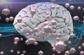
Swiss Study Shockingly Reveals That SARS-CoV-2 Triggered Inflammation Leads To Increased Neurotoxic Damage In Cortical Areas Of The Human Brain!
Source: Thailand Medical-NeuroCOVID Feb 20, 2022 3 years, 10 months, 1 week, 4 days, 2 hours, 59 minutes ago
NeuroCOVID: Researchers from the University Hospital Basel and University of Basel-Switzerland have in a new shockingly found that SARS-CoV-2 triggered inflammation leads to increased neurotoxic damage in cortical areas of the human brain!

To date, despite mounting evidence showing that the human host brain is a target of SARS-CoV-2, the consequences of the virus on the cortical regions of hospitalized patients and non-hospitalized patients are currently unknown.
The key aim of the research was to assess brain cortical gray matter volume (GMV), thickness (Th), and surface area (SA) characteristics in SARS-CoV-2 hospitalized patients with a wide range of neurological symptoms and their association with clinical indicators of inflammatory processes.
In all, a total of 33 patients were selected from a prospective, multicenter, cross-sectional study during the ongoing pandemic (August 2020-April 2021) at Basel University Hospital. Retrospectively biobank healthy controls with the same image protocol served as controls group.
For every anatomical T1w MPRAGE image, the Th and GMV segmentation were performed with the FreeSurfer-5.0. Cortical measures were compared between groups using a linear regression model. The covariates were age, gender, age*gender, MRI magnetic field strength, and total intracranial volume/mean Th/Total SA. The association between cortical features and laboratory variables was assessed using partial correlation adjusting for the same covariates. P-values were adjusted using false discovery rate (FDR).
The
NeuroCOVID study findings revealed a lower cortical gray matter volume in orbitofrontal and cingulate regions in patients compared to controls. The orbitofrontal grey matter volume was negatively associated with protein levels, CSF-blood/albumin ratio and CSF EN-RAGE level. CSF EN-RAGE and CSF/Blood-albumin ratio, which are neuroinflammatory biomarkers, were associated with cortical alterations in gray matter volume and thickness in frontal, orbitofrontal, and temporal regions.
The study findings confirm that viral-triggered inflammation leads to increased neurotoxic damage in some cortical areas.
The study findings were published on a preprint server and are currently being peer reviewed.
https://www.medrxiv.org/content/10.1101/2022.02.13.22270662v1
Numerous past studies have examined chronic neuronal dysfunction and hyperinflammatory responses in patients infected with severe acute respiratory syndrome coronavirus 2 (SARS-CoV-2). However, studies have not assessed these neurological symptoms and related neurological complications, observed in 80% of hospitalized COVID-19 cases.
The study team assessed several neurological symptoms and their linkage with clinical indicators of inflammatory processes in hospitalized COVID-19 patients.
The study team selected the Desikan-Killiany atlas, having 33 cortical regions per hemisphere, and studied its gray matter volume (GMV) (cm3), thickness (Th) (mm), and surface area (SA) (mm2) characteristics.
The team screened 33 patients at Basel University hospital who
participated in a multicenter, cross-sectional study between August 2020 and April 2021 and were 18 years or older. As controls, age- and sex-matched healthy individuals having the same image protocol were recruited from the Neurology Department, University of Basel.
The study team used a linear regression model to compare cortical measures between the study and the control groups.
The study team next performed high-resolution three-dimensional (3D) longitudinal relaxation time-weighted (T1w) magnetic resonance imaging (MRI) sequencing of the brain of all the eligible participants.
A subset of participants underwent cerebrospinal fluid (CSF) sampling. The laboratory tests included leukocytes, lactate, protein levels, CSF-blood/albumin-ratio, and five cytokines (plasma-tumor necrosis factor-related activation-induced cytokine (TRANCE), plasma-receptor for advanced glycation end-products binding protein (EN-RAGE), CSF-osteoprotegerin (OPG), CSF-TRANCE, and CSF-EN-RAGE).
The study team performed cytokines analysis using the Olink 96 target inflammation and neurology panels.
The study team employed the FreeSurfer-6.0 image analysis suite for the Th and GMV segmentation of each anatomical T1w magnetization-prepared rapid gradient-echo (MPRAGE) image, taken using MRI scanners. The study covariates were gender, age, age*gender, total intracranial volume/mean Th/total SA, and MRI magnetic field strength.
The team used partial correlation with adjustments to the same covariates to examine the potential links between cortical features and laboratory variables; further, they used false discovery rate (FDR) for p-values adjustments.
For the study, based on their neurological symptoms, there were three classes of study participants - class I, class II, class III, having mild, moderate, and severe neurological symptoms, respectively.
The study findings showed a lower GMV in cingulate and orbitofrontal cortical regions in some of the hospitalized SARS-CoV-2 patients; similar to previously reported multifocal MRI abnormalities in the hospitalized SARS-CoV-2 patients.
In details, a lower GMV was observed in the right rostral anterior cingulate, left medial orbitofrontal, and left superior frontal regions with patients-mean of 0.38, 0.84, and 3.75, respectively. Intriguingly, GMV was consistently negatively associated with protein levels, CSF/blood-albumin ratio, and CSF EN-RAGE.
It was found that after FDR correction, the patient cohort and the healthy controls showed no significant differences between Th and SA values. In the subset of patients considered for the CSF study, blood leukocytes were negatively associated with GMV in the right lateral orbitofrontal and left inferior temporal regions.
The study team said that as hypothesized, the brain alterations after SARS-CoV-2 infection is a neuroinflammatory response.
Co-researcher, Dr Cristina Granziera, a professor at the Department of Neurology, University Hospital Basel told
Thailand Medical News, “The study findings confirmed increased CSF levels of indirect inflammatory markers, including protein, blood/albumin ratio, and EN-RAGE.”
Interestingly, in 18 regions localized in the frontal, orbitofrontal, and temporal lobes of the brain cortex, the study team observed the highest number of negative correlations for protein levels. Of these proteins, CSF/blood/albumin-ratio and the EN-RAGE cytokine showed negative correlations in 15 and 17 cortical regions, respectively.
Importantly the CSF/blood/albumin-ratio showed a positive correlation with left rostral anterior cingulate, left lateral orbitofrontal, and right paracentral region of the brain cortex with p-values of 0.03, 0.02, and 0.04, respectively. But it negatively correlated with the left rostral anterior cingulate and right caudal middle frontal.
EN-RAGE showed a positive correlation with the left pars triangularis with a p-value of 0.002. Likewise, SA showed a significant correlation between the EN-RAGE and the right posterior cingulate cortex with a p-value of 0.04.
Th study findings showed that the relationship between ENRAGE, a cytokine that activates an inflammatory cascade, and CSF/blood-albumin ratio with increased volumes in some cortical areas suggested a SARS-CoV-2-triggered inflammatory process due to a secondary para-infectious complication or after a less probable direct invasion.
Furthermore, the study team noted a significant association between a decreased GMV and Th in the frontal, frontal-orbital, and temporal lobes of the brain’s cortex of hospitalized COVID-19 patients.
The study findings demonstrated an association between CSF inflammatory marker levels and GMV and Th changes in frontal, orbitofrontal, and temporal regions in hospitalized SARS-CoV-2 patients exhibiting different levels of neurological symptoms.
The study team suggested that their findings be confirmed and expanded in future longitudinal studies in larger cohorts.
It should be noted that many studies have also showed that the SARS-CoV-2 virus not only causes damage to the brain structures in hospitalized patients but also to those who were asymptomatic upon infection or merely had mild symptoms during infection!
https://www.thailandmedical.news/news/study-confirms-that-even-in-asymptomatic-to-mild-cases-of-infections,-sars-cov-2-leaves-a-mark-on-the-brain
https://www.thailandmedical.news/news/saudi-arabia-mri-study-provide-further-evidence-that-sars-cov-2-affects-the-brain-with-57-9-percent-of-severe-covid-19-patients-having-brain-lesions
https://www.thailandmedical.news/news/breaking-researchers-puzzled-as-new-study-shows-that-covid-19-patients-manifest-reduced-volume-of-brain-gray-matter
https://www.thailandmedical.news/news/study-indicates-that-even-those-with-mild-or-asymptomatic-covid-19-can-develop-brain-inflammation-and-other-neurological-conditions-in-long-term
For more about
SARS-CoV-2 And Brain Damages, keep on logging to Thailand Medical News.
