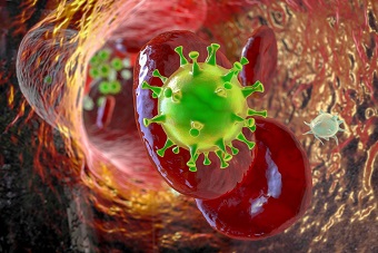
U.S. NIH Study Validates That SARS-CoV-2 Spike Proteins Causes Deformation Of Platelets And Irreversible Stochastic Activation Leading To Coagulopathies!
A new study by researchers from the National Heart, Lung, and Blood Institute, U.S. National Institutes of Health, United States using cellular cryo-electron tomography has clearly shown that the spike proteins of the SARS-CoV-2 virus causes deformation of platelets and also causes irreversible stochastic activation leading to coagulopathies.

The study also involved researchers from University of Oviedo-Spain and Osaka University Institute for Protein Research-Japan.
The SARS-CoV-2 coronavirus is highly pathogenicity due to its spike protein (S protein) contacting host-cell receptors.
A critical hallmark of COVID-19 is the occurrence of coagulopathies.
The study team reported the direct observation of the interactions between
SARS-CoV-2 Spike Proteins and platelets. Live imaging showed that the S protein triggers platelets to deform dynamically, in some cases, leading to their irreversible activation.
Detailed cellular cryo-electron tomography revealed dense decorations of S protein on the platelet surface, inducing filopodia formation. Hypothesizing that S protein binds to filopodia-inducing integrin receptors, the study team tested the binding to RGD motif-recognizing platelet integrins and found that the S protein recognizes integrin αvβ3.
The study findings infer that the stochastic activation of platelets is due to weak interactions of S protein with integrin, which can attribute to the pathogenesis of COVID-19 and the occurrence of rare but severe coagulopathies.
The study findings were published in the peer reviewed journal: Nature Communications.
https://www.nature.com/articles/s41467-023-36279-5
To date, SARS-CoV-2 has shown unique pathological symptoms that can lead to a wide range of coagulopathic events in severe cases.
The study team investigated the direct effect of S protein to the change in morphology of platelets at a molecular level, and the study directly visualized the binding of S protein to the platelet surface.
The study team hypothesized that the binding of the SARS-CoV-2 is mediated by integrin receptors based on the following reasons:
-the activation of platelets is governed by filopodia formation,
-filopodia formation is initiated by integrin receptors,
-the major receptors on the platelets are integrin receptors and
-SARS-CoV-2 S protein contains a RGD sequence in the RBD, which is recognized by a subtype of integrin, and therefore the study team tested the interaction of platelet-expressed integrins with S protein.
The study team’s integrin inhibition experiment using cilengitide and in vitro solid-phase binding assays support this hypothesis, particularly with the possibility that S protein recognizes integrin αvβ3.
It was found that the binding of S protein to integrin was much lower compared to the interaction of integrins with their physiological ligands, and interestingly, the study findings did not detect the binding to t
he major platelet integrin αIIbβ3.
In a past study, an increased binding of the activated integrin αIIbβ3 antibody PAC-1 to platelets was observed in the presence of S protein21.
https://jhoonline.biomedcentral.com/articles/10.1186/s13045-020-00954-7
Accordingly, this may be due to an inside-out effect, in which the outside-in signaling is activated by the direct binding of S protein to integrin αvβ3 and in turn, αIIbβ3 would get activated through the intracellular signaling (inside-out).
The study team surmise that the weak affinity of S protein to platelet integrin receptors and the reversible binding, may reflect the fact that blood clotting defects observed in patients are rare complications and occur in severe cases of COVID-19.
However, here it should also be noted that there are other receptors on platelets that may also be accountable for the interaction with S protein33,53 and combinatory effects of the binding of S protein to multiple receptors may also occur.
https://www.nature.com/articles/s41392-020-00426-x
https://ashpublications.org/blood/article/120/15/e73/30645/The-first-comprehensive-and-quantitative-analysis
It has been noted that SARS-CoV-2 can be found in the blood stream of COVID-19 patients, and an open question is how it can lead to rare but severe coagulation defects.
https://translational-medicine.biomedcentral.com/articles/10.1186/s12967-020-02693-2
The study findings showed that the deformation of platelets itself does not always alter their intracellular signaling, or induces activation.
Rather, it appears that platelets exposed to S protein are primed for the activation upon further stimuli, such as the attachment to an adhesion surface.
From such an observation, the study team speculates that the combination of the direct binding of S protein to platelets and other identified coagulation factors may induce a synergistic and irreversible activation of platelets, leading to coagulation.
It has been found that during SARS-CoV-2 infection, several other procoagulant players are active, for example the formation of neutrophil extracellular traps, the release of TF, elevated fibrinogen levels and dysregulated release of cytokines, creating a hypercoagulative environment in the context of COVID-19.
https://ashpublications.org/blood/article/136/10/1169/461219/Neutrophil-extracellular-traps-contribute-to
https://www.nejm.org/doi/full/10.1056/NEJMra2026131
https://www.sciencedirect.com/science/article/pii/S0268960X20301119
https://www.nejm.org/doi/full/10.1056/NEJMra2026131
The study team visualized the adaptable attachment of S protein to the platelet plasma membrane with a high degree of flexibility for the engagement to continuously curved membrane surfaces. Similarly, it has been reported that the stalk domain of S protein proximal to the viral membrane surface contains three hinges, presumably allowing the flexible motion of individual S protein on the viral surface to adapt to curved host cell surfaces.
https://www.science.org/doi/full/10.1126/science.abd5223
This dual flexibility likely increases the probability for S protein to attach to a host cell receptor, thus, allowing an efficient action of S protein to the membrane surface.
For the latest on
SARS-CoV-2 Spike Proteins, keep on logging to Thailand Medical News.
