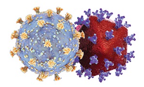
University Of Kansas Study Shockingly Reveals That The Envelope Protein Of SARS-Cov-2 Potently Inhibits HIV-1 Infection!
Source: SARS-CoV-2 - HIV Mar 30, 2022 3 years, 3 weeks, 6 days, 5 hours, 5 minutes ago
A new study by researchers from University of Kansas Medical Center-USA has found that the envelope protein of SARS-Cov-2 potently inhibits HIV-1 infection!

It has been found that the human coronavirus SARS-CoV-2 encodes for a small 75 amino acid transmembrane proteins known collectively as the envelope (E) protein.
The E protein forms an ion channel, like the viroporins from human immunodeficiency virus type 1 (HIV-1) (Vpu) and influenza A virus (M2).
The E protein of coronaviruses is a viroporin that is required for efficient release of infectious virus and for viral pathogenicity.
The study team analyzed HIV-1 virus infectivity in the presence of four different β-coronavirus E proteins.
The study findings showed SARS-CoV-2 and SARS-CoV E proteins reduced HIV-1 yields by approximately 100-fold while MERS-CoV or HCoV-OC43 E proteins restricted HIV-1 infectivity to a lesser extent. This was also reflected in the levels of HIV-1 protein synthesis in cells.
The study findings showed mechanistically that that the E protein neither affected reverse transcription nor genome integration.
However,
SARS-CoV-2 E protein activated the ER-stress pathway associated with the phosphorylation of eIF-2α, which is known to attenuate protein synthesis in cells.
The study findings showed the various E proteins and the SARS-CoV-2 N protein did not significantly down-regulate bone marrow stromal cell antigen 2 (BST-2) while the spike (S) proteins of SARS-CoV and SARS-CoV-2, and HIV-1 Vpu efficiently down-regulated cell surface BST-2 expression.
The study findings show for the first time that the E protein also triggers autophagy and that viroporin from a coronavirus can restrict infection of another virus such as HIV-1.
The study findings were published on a preprint server and are currently being peer reviewed.
https://www.biorxiv.org/content/10.1101/2022.03.23.485576v1
The SARS-CoV-2 virus, a highly transmissible human CoV (HCoV), emerged in late 2019 and caused the ongoing coronavirus disease 2019 (COVID-19) pandemic. The unprecedented morbidity and mortality caused by COVID-19 have underlined the critical need to learn more about the role of viral proteins in virus pathogenesis and replication.
It has been found that the E protein is a small 75 amino acid transmembrane protein encoded by the human CoVs, including SARS-CoV-2. The CoVs E protein, similar to the viroporins from the influenza A virus, i.e., matrix protein 2 (M2), and HIV-1, i.e., viral protein U (Vpu), creates an ion channel. The CoVs E protein is essential for viral pathogenicity and the successful release of infectious viruses into the host.
The study team evaluated the infectivity of the HIV-1 virus in the vicinity of four distinct β-CoV E proteins. The team analyzed the biological characteristics of four β-CoVs (SARS-CoV-2, SARS-CoV, Middle East respiratory CoV (MERS-CoV), and HCoV-OC43) E proteins in the presence of HIV-1.
For the study, vectors encoding the HIV-1 genome and CoVs proteins were transfected into human embryonic kidney 293 (HEK293) cells. HIV-1 in
fectivity was quantified using the TZM-bl cell line (derived from Henrietta Lacks cell clone).
The U.S. National Institutes of Health (NIH) HIV Reagent Branch of America provided plasmids encompassing the complete HIV-1 NL4-3 genome, i.e., pNL4-3 and pNL4-3ΔVpu. Immunofluorescence investigations of COS-7 cell (African green monkey kidney fibroblast-like cell line) transfections were used to determine SARS-COV-2 E protein intracellular localization.
The study findings showed that co-transfection of HEK293 cells with vectors encoding the SARS-CoV-2 E protein and plasmids containing either the HIV-1ΔVpu or HIV-1 (strain NL4-3) genomes significantly limited the infectious HIV-1 release; similar to the herpes simplex virus 1 (HSV-1) glycoprotein D (gD) protein.
Immunoprecipitation of SARS-CoV2 E proteins and gD from cell lysates demonstrated that these proteins were expressed efficiently during co-transfections.
It was also found that SARS-CoV-2 E protein did not affect the synthesis of infectious HSV-1 progeny virus 24 and 48 hours following the infection.
It was also found that similar to the SARS-CoV-2 E protein, the MERS-CoV, SARS-CoV, and HCoV-OC43 E proteins were largely localized in the Golgi apparatus and endoplasmic reticulum (ER) cell regions, with no expression at the cell surface.
Most importantly, the study findings showed that SARS-CoV and SARS-CoV-2 E proteins lowered HIV-1 production by nearly 100-fold.
However, HCoV-OC43 or MERS-CoV E proteins inhibited HIV-1 infectivity to a limited extent. Replacing the significantly conserved proline within the SARS-CoV-2 E's cytoplasmic domain abolished the limitation on HIV-1 infection.
The study findings also showed that the presence of SARS-CoV and SARS-CoV-2 E proteins lowered the levels of HIV-1 proteins. By contrast, the HCoV-OC43 and MERS-CoV E proteins only reduced the HIV-1 protein synthesis moderately. The CoVs E proteins did not affect genome integration or reverse transcription of the HIV-1 virus. Nonetheless, via phosphorylating eukaryotic initiation factor 2α (eIF-2α), the SARS-CoV and SARS-CoV-2 E proteins stimulated the ER-stress pathway, which reduces protein production in cells.
The study findings demonstrated that the SARS-CoV-2-induced restriction of HIV-1 infection was due to its E protein's capacity to stimulate eIF-2α phosphorylation.
It was also found that the four CoVs E protein or SARS-CoV-2 nucleocapsid (N) proteins did not down-modulate the cell surface expression of bone marrow stromal cell antigen 2 (BST-2). Nevertheless, the HIV-1 Vpu protein and SARS-CoV-2 and SARS-CoV spike (S) proteins drastically down-regulated the BST-2 cell surface expression.
Accompanying studies indicated that the CoVs E proteins triggered autophagy in HIV-1.
To date, this was the first study revealing that viroporins from a heterologous virus, i.e., CoVs E proteins, could reduce HIV-1 infection.
The research data showed that CoVs such as SARS-CoV-2 and SARS-CoV significantly limited HIV-1 infection, whereas HCoV-OC43 and MERS-CoV were less restrictive.
Also, similar observations were seen in the amounts of HIV-1 protein production in cells. The SARS-CoV-2 E protein, instead of interfering with RNA synthesis or viral integration, lowered viral protein production. The CoVs E protein-induced ER stress leads to phosphorylation of eIF-2, which inhibits protein synthesis of HIV-1.
Most importantly, the study findings showed that E proteins of four CoVs and the SARS-CoV-2 N protein did not substantially lower BST-2 expression. On the contrary, the HIV-1 Vpu, SARS-CoV-2, and SARS-CoV S proteins decreased BST-2 cell surface expression.
The study team proposed that further detailed studies are warranted to understand the CoVs S protein domains relevant for their interactions with BST-1.
Before any wild Westerners start going out and having unprotected sex while they are infected with the SARS-CoV-2 virus, assuming that they will now not contract HIV, please kindly do more due diligence about the effects of Long COVID and what you could be passing to your partners and also yourself or worst, possibly give rise to coinfections and new recombinant variants if both of you or the rest of you (depending what you are all into?) are infected with different variants! (Am sure that Robert Hunter Biden will be thrilled by this new discovery!)
For more on
SARS-CoV-2 And HIV, keep on logging to Thailand Medical News.
