University of North Carolina Murine Study Shows That SARS-CoV-2 Infections Ultimately Lead To Chronic Pulmonary Epithelial And Immune Cell Dysfunction!
Source: Long COVID or PASC Jul 09, 2022 2 years, 9 months, 6 days, 17 hours, 19 minutes ago
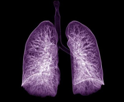
A new murine study by researchers from the University of North Carolina at White Chapel-USA has found that SARS-CoV-2 infections ultimately lead to chronic pulmonary epithelial and immune cell dysfunction, including the development of fibrosis.

It is already known that a subset of individuals who recover from COVID-19 disease develop what is known as
Long COVID or post-acute sequelae of SARS-CoV-2 (PASC).
However, the mechanistic basis of PASC-associated lung abnormalities suffers from a lack of longitudinal tissue samples. The mouse-adapted severe acute respiratory syndrome coronavirus 2 (SARS-CoV-2) strain MA10 produces an acute respiratory distress syndrome (ARDS) in mice similar to humans.
In order to investigate PASC pathogenesis, studies of MA10-infected mice were extended from acute to clinical recovery phases. At 15 to 120 days post-virus clearance, pulmonary histologic findings included subpleural lesions composed of collagen, proliferative fibroblasts, and chronic inflammation, including tertiary lymphoid structures.
Detailed longitudinal spatial transcriptional profiling identified global reparative and fibrotic pathways dysregulated in diseased regions, similar to human COVID-19. Populations of alveolar intermediate cells, coupled with focal up-regulation of pro-fibrotic markers, were identified in persistently diseased regions.
In the study, early intervention with the antiviral EIDD-2801 reduced chronic disease, and early anti-fibrotic agent (nintedanib) intervention modified early disease severity. This murine model provides opportunities to identify pathways associated with persistent SARS-CoV-2 pulmonary disease and test countermeasures to ameliorate PASC.
The study findings were published in the peer reviewed journal: Science Translational Medicine
https://www.science.org/doi/10.1126/scitranslmed.abo5070
It is already known that SARS-CoV-2 infection causes Acute Lung Injury or ALI, Acute Respiratory Disease or ARDS, and PASC. Although PASC encompasses non-respiratory sequelae, including cardiovascular and neurologic disease, pulmonary manifestations are especially common, including
Community-acquired pneumonia or CAP and Pulmonary Fibrosis or PF.
Computed tomography or CT scans reveal chronic COVID-19 pulmonary findings as evidenced by ground glass opacities (44%) and fibrosis (21%) after acute COVID-19 infection and fibrotic-like changes (35%) 6 months after severe human COVID-19 pneumonia.
https://pubmed.ncbi.nlm.nih.gov/34278556/
https://pubmed.ncbi.nlm.nih.gov/33497317/
Past pathology studies of COVID-19 lungs obtained at autopsy reveal similar late findings, such as CAP and PF.
https://pubmed.ncbi.nlm.nih.gov/34841234/
https://pubmed.ncbi.nlm.nih.gov/32989525/
ttps://pubmed.ncbi.nlm.nih.gov/32926596/">https://pubmed.ncbi.nlm.nih.gov/32926596/
The study team focused their studies of PASC in the SARS-CoV-2 MA10 mouse model of COVID-19 and specifically on the pulmonary features of PASC.
To date, the understanding of PASC and COVID-19-induced CAP and PF is poor, and countermeasures are limited due to the wide spectrum of potential disease pathophysiologies. Though better studied, these limitations are also observed in infections with other viral respiratory pathogens such as influenza.
Hence, human and animal model data comparing the chronic sequelae of a spectrum of respiratory viruses should, therefore, identify unique versus shared disease manifestations and mechanisms of disease that will contribute to not only knowledge of COVID-19 PASC, but virus infection-mediated sequelae of the lung in general.
A recent study involving a chronic (30 dpi) SARS-CoV-2 infection model was reported in immunosuppressed, humanized mice characterized by persistent virus replication and chronic inflammation with fibrotic markers, typical of rare infections seen in immunosuppressed humans who cannot clear virus.
https://pubmed.ncbi.nlm.nih.gov/34921308/
The study team in contrast, reports a mouse model of long-term pulmonary sequelae of SARS-CoV-2 infection that persisted after virus clearance and was more characteristic of disease outcomes seen at the general patient population.
Alarmingly, in the SARS-CoV-2 MA10 model, surviving older mice cleared infection by 15 dpi but exhibited damaged pulmonary epithelia accompanied by secretion of a spectrum of pro-inflammatory and pro-fibrotic cytokines often up-regulated in fibrotic disease in humans, including IL-1β, TNF-α, GM-CSF, TGF-β, IL-33, and IL-17A.
https://err.ersjournals.com/content/17/109/151.short
Similar to humans, surviving SARS-CoV-2-infected mice developed heterogeneous, persistent pulmonary lesions of varying severity by 30 to 120 dpi, presenting with abnormally repairing AT2 cells, interstitial macrophage and lymphoid cell accumulation, myofibroblast proliferation, and interstitial collagen deposition, particularly in subpleural regions.
https://pubmed.ncbi.nlm.nih.gov/25383540/
https://pubmed.ncbi.nlm.nih.gov/30209189/
Heterogeneous subpleural opacities and fibrosis were detected by Micro-CT in surviving mice, similar to human studies.
https://pubmed.ncbi.nlm.nih.gov/34278556/
Though most acute cytokine concentrations returned to normal values by 30 dpi, DSP and RNA-ISH data revealed focally prolonged up-regulation of cytokine signaling, including TGF-β, in sub-pleural fibrotic regions. Importantly, similar heterogeneous cellular and fibrotic features in subpleural regions are also evident in patients with late stage COVID-19.
SARS-CoV-2 MA10 infection principally caused acute loss of distal airway club cell (Scgb1a1) and alveoli AT2 cell (Sftpc) marker expression, phenotypes consistent with SARS-CoV-2 cellular tropism in humans. The expression of club and AT2 cell genes were variably restored by 15 dpi, as demonstrated by DSP and RNA-ISH data.
The study team speculates that a key variable determining the ability of the alveolar region to repair, or not, reflects the capacity of surviving or residual AT2 cells to regenerate an intact alveolar epithelium.
Importantly the failure of AT2 cells to replenish themselves or AT1 cells and repair alveolar surfaces in subpleural regions may reflect the intensity of SARS-CoV-2 infection.
From past data obtained via COVID-19 autopsy lungs, an accumulation of replication-defective and pro-inflammatory (ADI/DATP/PATS) transitional cells emerge early after SARS-CoV-2 infection and may persist, associated with continued inflammation and failure of repair.
https://pubmed.ncbi.nlm.nih.gov/33915569/
The study team’s longitudinal mouse model data support this notion as evidenced by the observation that ADI/DATP/PATS cells were detected at 2 dpi and persisted through 30 dpi in diseased, but not morphologically intact, alveolar regions. These ADI/DATP/PATS cells were notable for up-regulation of pathways associated with senescence, Hif1α, and pro-inflammatory cytokines such as IL-1β, consistent with low cycling rates, a failure to replenish AT2 and AT1 cells, and a pro-inflammatory phenotype.
As evidenced by the return of Sftpc expression by 15 dpi in intact alveolar regions, a fraction of the ADI/DATP/PATS cells likely regenerated mature Sftpc-expressing AT2 cells. Notably, the study team’s longitudinal studies revealed that the gene expression profiles of ADI/DATP/PATS cells are dynamic over the evolution of lung disease.
.jpg) Digital spatial profiling reveals distinct transcriptional pathway changes during acute and late stages of SARS-CoV-2 disease. (A and B) DSP heatmaps are shown for differentially expressed genes (DEGs) in ROIs across all time points in (A) distal airway and (B) alveolar tissue compartments. DEGs were obtained by comparing DSP Q3 normalized counts of transcripts between ROIs at 2, 15, and 30 dpi versus mock-infected 1-year-old female BALB/c mice. (C) Normalized enrichment scores (ES) and adjusted p-values obtained by DSP pathway enrichment analysis are shown for distal airway and alveolar ROIs at 2, 15, or 30 dpi versus mock. Statistical analyses used are detailed in methods. NS, not significant.
Digital spatial profiling reveals distinct transcriptional pathway changes during acute and late stages of SARS-CoV-2 disease. (A and B) DSP heatmaps are shown for differentially expressed genes (DEGs) in ROIs across all time points in (A) distal airway and (B) alveolar tissue compartments. DEGs were obtained by comparing DSP Q3 normalized counts of transcripts between ROIs at 2, 15, and 30 dpi versus mock-infected 1-year-old female BALB/c mice. (C) Normalized enrichment scores (ES) and adjusted p-values obtained by DSP pathway enrichment analysis are shown for distal airway and alveolar ROIs at 2, 15, or 30 dpi versus mock. Statistical analyses used are detailed in methods. NS, not significant.
Similar to humans, CD4+ and CD8+ T cell populations increased in SARS-CoV-2-diseased areas of mouse lungs, and peripheral lymphoid aggregations were a feature of chronic disease. These features were consistent across all analyses, including immunohistochemistry, DSP, and flow cytometry data.
A significant macrophage feature, identified by DSP and flow cytometry data, was expansion of the interstitial macrophage population, consistent with human data.
Interestingly, the subpleural regions exhibited the most striking histologic evidence of immunologic cell recruitment and activation of adaptive immune, hypoxia, fibrotic, and extracellular matrix pathways in association with ADI/DATP/PATS cells.
Clues to the etiology of the late-stage alveolar CAP/PF response emerged from comparisons to infection in bronchioles. The alveolar regions exhibited persistent CAP/PF disease, particularly in subpleural regions. This finding is consistent with proposed relationships between the maximal pulmonary mechanical stretch imposed on the subpleural region during tidal breathing and activation of stretch-induced fibrotic pathways, such as TGF-β-mediated signaling, during periods of injury.
In contrast, despite similar infection, bronchioles repaired without evidence of organizing or fibrotic sequelae. Bronchioles may be protected from this adverse fate by tissue-specific ISG responses to control the duration or severity of infection. In this context, several ISGs, including Ifitm1 and Ifitm2, exhibited clear differences in tissue specific expression or persistence through 30 dpi.
Other possible relevant variables that may favor bronchiolar repair include: 1) more “controlled” cell death, such as apoptosis; 2) a less damaged basement membrane architecture; and 3) inability of club cells to enter an intermediate, ADI/DATP/PATS cell equivalent.
Murine models of acute and chronic viral disease are critical also for countermeasure development.
Though speculative, early direct-acting antiviral treatment may forestall chronic lung and other organ PASC manifestations.
From data of preclinical studies of anti-fibrotic agents in reducing the severity of PF responses to chemical agents, the study team tested the concept that early intervention with an anti-fibrotic agent may reduce the severity of PF following SARS-CoV-2 infection.
https://pubmed.ncbi.nlm.nih.gov/25745043/
The drug, nintedanib was administered from 7 dpi blunted maximal fibrotic responses to virus at 15 dpi, supporting the concept that early intervention with anti-fibrotic agents may attenuate post-SARS-CoV-2 severe disease trajectories.
This finding suggests that early administration of direct-acting antivirals or antifibrotic drugs may help reduce human pulmonary fibrosis, and combination therapies may further increase efficacy and prognoses.
Further studies of other anti-fibrotic candidates and host immune modulators will be important in continuing to develop PASC treatments. COVID-19 in mice and humans represent key findings that may prove translatable to other future emerging coronavirus disease pathologies. Moreover, comparative models of viral induced chronic lung disease are needed to identify common and unique pathways associated with virus-induced CAP and are key for the development of new therapeutic options for treating PASC.
The study findings show that acute and chronic disease phases in SARS-CoV-2 MA10-infected mice strongly recapitulate the pulmonary pathology observed in patients with COVID-19 and provides an excellent model for studies of pathogenesis and selected countermeasures. Transgenic and vectored expression of human ACE2 mouse models are also commonly used to understand SARS-CoV-2 pathogenesis, but studies investigating the long-term pulmonary effects of infection in these models have not been reported.
The research findings provide new data for modeling chronic SARS-CoV-2 and indeed other respiratory viral pathogens. By extending the studies of SARS-CoV-2 MA10 sequelae in mice out to 120 days in 1-year-old BALB/c mice, the study team observed that many chronic phenotypes first observed at 15 dpi were maintained for the entire 120 d observational period, extending from acute to ongoing to chronic COVID-19 defined disease classifications used in human populations.
Importantly, the observation that fibrotic pulmonary disease in young BALB/c and aged C57BL/6J mice peaked at 15 dpi, but waned by 30 dpi in many animals compared to aged BALB/c mice suggests that multiple time points for extended time intervals will be required for informative studies across viral pathogens.
In conclusion, the SARS-CoV-2 MA10 mouse model provides opportunities to longitudinally study the molecular mechanisms and pathways mediating long-term COVID-19 pulmonary sequelae as relates to human PASC and to evaluate treatments.
Future studies will be needed to determine if other chronic, extrapulmonary organ sequelae develop after acute infection, the current model supports high-priority research directions that include SARS-CoV-2 infection of transgenic lineage tracing reporter mice to define longitudinally the fates of infected club and AT2 cells, ADI/DATP/PATS cell transitions, mechanisms of cell death, and epithelial cell regeneration and repopulation following infection.
The findings also provide the foundation for understanding the role of sex, host genetics, and immunological contributions, through knockout and Collaborative Cross studies, in defining PASC outcomes.
Further studies should be feasible in this model to investigate the non-pulmonary sequelae of PASC, including cardiovascular, neurological, and behavior manifestations in mice.
For the latest
COVID-19 Research, keep on logging to Thailand
Medical News.

.jpg) Digital spatial profiling reveals distinct transcriptional pathway changes during acute and late stages of SARS-CoV-2 disease. (A and B) DSP heatmaps are shown for differentially expressed genes (DEGs) in ROIs across all time points in (A) distal airway and (B) alveolar tissue compartments. DEGs were obtained by comparing DSP Q3 normalized counts of transcripts between ROIs at 2, 15, and 30 dpi versus mock-infected 1-year-old female BALB/c mice. (C) Normalized enrichment scores (ES) and adjusted p-values obtained by DSP pathway enrichment analysis are shown for distal airway and alveolar ROIs at 2, 15, or 30 dpi versus mock. Statistical analyses used are detailed in methods. NS, not significant.
Digital spatial profiling reveals distinct transcriptional pathway changes during acute and late stages of SARS-CoV-2 disease. (A and B) DSP heatmaps are shown for differentially expressed genes (DEGs) in ROIs across all time points in (A) distal airway and (B) alveolar tissue compartments. DEGs were obtained by comparing DSP Q3 normalized counts of transcripts between ROIs at 2, 15, and 30 dpi versus mock-infected 1-year-old female BALB/c mice. (C) Normalized enrichment scores (ES) and adjusted p-values obtained by DSP pathway enrichment analysis are shown for distal airway and alveolar ROIs at 2, 15, or 30 dpi versus mock. Statistical analyses used are detailed in methods. NS, not significant.