For All The Latest Medical News, Health News, Research News, COVID-19 News, Dengue News, Glaucoma News, Diabetes News, Herb News, Phytochemical News, Cardiology News, Epigenetic News, Cancer News, Doctor News, Hospital News
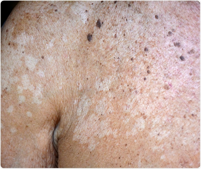
Idiopathic guttate hypomelanosis (IGH) is a common benign skin condition. It appears as multiple round or oval macules of depigmentation or hypopigmentation, mostly over the upper or lower extremities.
Many other conditions resemble this dermatosis, and the more common ones are discussed briefly in the following section. The lesions in IGH are of a characteristic size and the distribution, age of onset, smooth surface without fine scaling, and negative history of dermatitis or viral infection all point to this condition rather than any other.
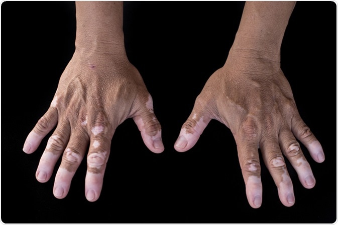
Vitiligo is a dermatosis marked by the appearance of white patches on various parts of the body due to atrophy of the melanocytes in that region of skin. It may affect the mucous membranes inside the mouth or nose, as well as the skin. While the exact etiology is as yet unknown, it is thought to be related to autoimmunity. The patches occur most commonly over sun-exposed areas such as the hands or feet, arms and face, and are often associated with early graying of hair as well. The patches may spread or remain stationary in various individuals, and their extension is sometimes associated with stressful periods of life in some individuals.

Pityriasis versicolor is a common yeast infection caused by fungi belonging to the genus Malassezia, which are part of the normal microbial flora of the human body. The affected skin appears to flake off and is characterized by discolored patches, mostly over the chest and the back. It is also sometimes called Tinea versicolor though this is not strictly accurate. It is a disease of young adults but may also occur in younger age groups. The ‘versicolor’ part of the name refers to the varied colors of the patches, from coppery to pink or even hypopigmented, the latter especially occurring in dark-skinned individuals. It may cause mild pruritus but is most commonly asymptomatic.

Pityriasis alba is also a dermatosis characterized by scaling patches, but these are reddish at first, finally turning pale. The patches are round or oval, and often occur in multiples on the face and arms. It is associated with atopic dermatitis and most often occurs in young children, resolving by the time they grow into adulthood. At most, moisturizing creams or sometimes a corticosteroid cream of low potency may be recommended by way of treatment.
Tuberous sclerosis is a rare condition of genetic origin that manifests as benign tumors in many parts of the body, producing signs and symptoms that vary with the location of the growths. Hypopigmented or thickened skin patches may occur as part of this syndrome. Acneform lesions are also common.
Lichen sclerosus is a rare condition of the skin that results in whitish patches of fragile or wrinkled skin over various parts of the body. The most common sites are the vulval, foreskin or perianal regions, with the highest incidence in perimenopausal women. The lesions are pruritic or painful, and may bleed or blister in severe cases, even causing the skin to tear. Dyspareunia is a troublesome complication. It resolves spontaneously in a few cases but in most patients it needs to be treated.
Some warts may resolve with hypopigmentation especially after the use of some home remedies or harsh chemicals which cause melanocyte regression.
Some inflammatory lesions such as eczema or seborrheic dermatitis, discoid lupus or lichen planus, or following skin trauma, as with burns, laser therapy or cryotherapy, result in hypopigmentation of the affected region because of decreased melanocyte function. The skin patches are lighter than the surrounding skin but there are no scales unless associated with scaling dermatoses such as psoriasis or eczema. The patches are flat, and are more noticeable in dark-skinned individuals. The clinical appearance is usually characteristic along with the history.