For All The Latest Medical News, Health News, Research News, COVID-19 News, Dengue News, Glaucoma News, Diabetes News, Herb News, Phytochemical News, Cardiology News, Epigenetic News, Cancer News, Doctor News, Hospital News
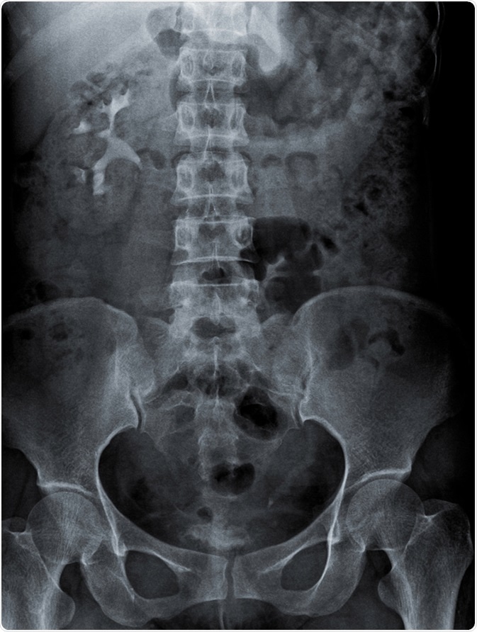
KUB stands for kidney, ureter and bladder. A KUB radiograph is an X-ray performed for the purpose of examining the urinary system and its surrounding structures.
The region covered by a KUB radiograph includes the area that spans the superior poles of the kidneys downwards to the pubic symphysis. This radiograph, like all X-rays, employs the use of external electromagnetic beams to acquire images of internal tissues and organs.
These beams pass through our tissues and project onto plates that are treated beforehand to create a negative (i.e. a picture).

Patients presenting with acute undiagnosed abdominopelvic pain, which may or may not be accompanied by nausea, vomiting, diarrhea and/or voiding irregularities, usually have a KUB radiograph ordered as an initial diagnostic test.
The symptoms suffered by these patients may have a myriad of etiological conditions, such as urinary system stones, gallstones, malignancies and gastrointestinal blockage.
Acute pathologies are not the only indications for this test, as KUB radiographs may also be necessary for other medical purposes, such as confirming the correct placement of stents and feeding tubes within the abdomen.
No special patient preparation is necessary before undergoing a KUB radiographic procedure. Radio-opaque objects such as some types of clothing, jewelry, accessories and metals may need to be removed as these interfere with the quality of the images.
Medications that may have been taken in the days prior to the procedure need to be communicated to the physician as these may also have an effect on the image quality.
Where applicable and possible, patients may be asked to empty their bladders beforehand. Pregnancy needs to be ruled out, to avoid exposing the developing child to unnecessary radiation.
At the moment the X ray is being taken, the patient may be asked to hold the breath briefly. This helps to avoid respiratory movement which might blur the image and result in an image that is not clear.
The patient might be positioned in different ways for the X-ray depending on suspected pathology.
The entire process of taking the X-ray is painless, but the positioning during the procedure might not be comfortable for some people, especially if they are having an acutely painful condition.
Following the procedure, a radiologist examines the images and makes a report, which is sent to the physician attending to the patient.
Although KUB radiography is a fast, non-invasive and inexpensive procedure, there are some risks involved. The most important risk factor is the possibility of cancer associated with radiation exposure.
However, this risk is usually cumulative and depends on the exposure to radiation over one’s lifetime. Secondly, the dosages used for KUB radiography are small and no radiation remains in the person’s body once the procedure is completed.
Furthermore, the benefits of diagnosing and treating the acute medical condition, which could be otherwise associated with direct morbidity and mortality, outweigh the hypothetical risk.